Neuroanatomy S1 E1: Intro to the Central Nervous System #neuroanatomy #science #medicine #brain
Summary
TLDRThis informative video explores the intricate structure and functions of the human brain, highlighting its weight, complexity, and essential role in defining human individuality. With approximately 86 billion neurons forming trillions of synapses, the brain is compared to the Milky Way in its vastness. Key anatomical regions, including the forebrain, brainstem, and cerebellum, are examined, alongside the significance of the ventricular system filled with cerebrospinal fluid. The presentation also covers brain imaging techniques and the distinction between gray and white matter, offering a comprehensive understanding of this remarkable organ.
Takeaways
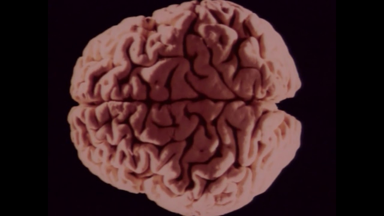
- 🧠 The adult human brain weighs approximately 1.5 kilograms, making up less than 2% of body weight.
- 🌌 The brain contains around 86 billion neurons, each forming an average of 7,000 connections, leading to 100 to 500 trillion synapses.
- 🧩 The number of neurons in the human brain is comparable to the number of stars in the Milky Way, highlighting its complexity.
- 🔍 The brain can be divided into anatomical and functional regions, beginning with the forebrain, which includes the cerebral hemispheres and diencephalon.
- 🔗 The corpus callosum connects the right and left hemispheres, consisting of approximately 200 to 250 million projections.
- 📏 The brain is highly folded to fit within the skull, creating gyri (ridges) and sulci (furrows), which increase surface area.
- 💧 The ventricular system of the brain is filled with cerebrospinal fluid (CSF) and includes lateral, third, and fourth ventricles, connected by the cerebral aqueduct.
- 🔄 Different planes (coronal, axial, sagittal) are used to section the brain for imaging and anatomical studies.
- ⚪ Gray matter consists of nerve cell bodies and can be found in the cortex and deep nuclei, while white matter consists of myelinated axons.
- 🌐 Understanding brain anatomy is essential for comprehending its complex functions, including language, thought, and memory.
Q & A
What is the approximate weight of the adult human brain?
-The adult human brain weighs approximately one and a half kilograms.
What percentage of the body's weight does the brain represent?
-The brain makes up less than 2% of a person's body weight.
What key functions does the brain perform?
-The brain is responsible for generating language and thought, attention, consciousness, memory, and imagination.
Why is the brain highly folded?
-The brain is highly folded to fit into the skull and accommodate the massive number of neurons and connections needed.
How many neurons are estimated to be in the human brain?
-There are an estimated 86 billion neurons in the human brain.
What is the significance of the corpus callosum?
-The corpus callosum is the largest white matter tract in the brain, connecting the right and left hemispheres with an estimated 200 to 250 million projections.
What anatomical regions make up the forebrain?
-The forebrain is composed of the cerebral hemispheres and deep structures, including the thalamus and hypothalamus.
What are the three parts of the brainstem?
-The three parts of the brainstem are the midbrain, pons, and medulla.
What is the role of cerebrospinal fluid (CSF) in the brain?
-Cerebrospinal fluid fills the space deep within the brain and helps protect and cushion the brain and spinal cord.
What are gyri and sulci in the context of the brain's structure?
-Gyri are the ridges on the brain's surface, while sulci are the furrows or grooves that separate them.
Outlines

This section is available to paid users only. Please upgrade to access this part.
Upgrade NowMindmap

This section is available to paid users only. Please upgrade to access this part.
Upgrade NowKeywords

This section is available to paid users only. Please upgrade to access this part.
Upgrade NowHighlights

This section is available to paid users only. Please upgrade to access this part.
Upgrade NowTranscripts

This section is available to paid users only. Please upgrade to access this part.
Upgrade NowBrowse More Related Video

Erase Una Vez... El Cuerpo Humano - El cerebro

AP Psychology- The Human Brain

Sistem Saraf: Otak Manusia | Ilmu Biomedik Dasar | Brainy Panda

What is Brain ? Exploring the Brain's Structure and Functions
The Brain | Part 13 | Discovery Channel Body Atlas
Nervous System - Get to know our nervous system a bit closer, how does it works? | Neurology
5.0 / 5 (0 votes)