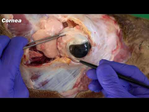
Extraocular Muscles | Eye Anatomy
Summary
TLDRIn this tutorial by Peter from Anatomy Zone, viewers learn about the seven extraocular muscles that control eye movement and eyelid elevation. These muscles are categorized into two groups: the levator palpebrae superioris for eyelid movement, and six muscles responsible for eye motion, including the superior rectus, inferior rectus, medial rectus, lateral rectus, superior oblique, and inferior oblique. Each muscle's origin, insertion, and innervation are discussed, highlighting their roles in coordinated eye movements such as elevation, depression, abduction, and adduction. The video emphasizes the collaborative function of these muscles, enhancing the viewer's understanding of ocular anatomy.
Takeaways
- 😀 The extraocular muscles are located in the orbit and control the movements of the eyeball and the superior eyelid.
- 🧑⚕️ There are seven extraocular muscles: levator palpebrae superioris, superior rectus, inferior rectus, medial rectus, lateral rectus, inferior oblique, and superior oblique.
- 🔍 The extraocular muscles can be categorized into two groups: those responsible for eye movement (recti and oblique muscles) and those responsible for eyelid movement (levator palpebrae superioris).
- ⬆️ Eye movements include elevation (upward movement), depression (downward movement), abduction (lateral movement), adduction (medial movement), and torsion (rotation).
- 👁️ The levator palpebrae superioris is the only muscle responsible for raising the upper eyelid and is innervated by the oculomotor nerve (Cranial Nerve III).
- 🪢 The four recti muscles (superior, inferior, medial, lateral) originate from the common tendinous ring and attach to the sclera of the eyeball.
- 📉 The superior rectus muscle elevates and medially rotates the eyeball, while the inferior rectus muscle depresses and laterally rotates it, both innervated by the oculomotor nerve.
- 👉 The lateral rectus muscle is responsible for abduction and is innervated by the abducens nerve (Cranial Nerve VI).
- 🔄 The oblique muscles (superior and inferior) have an angular approach to the eyeball, with the superior oblique innervated by the trochlear nerve (Cranial Nerve IV) and the inferior oblique by the oculomotor nerve.
- 🤝 All extraocular muscles work together to coordinate eye movements, ensuring smooth and accurate positioning of the eyeball.
Q & A
What are the extraocular muscles?
-The extraocular muscles are a group of seven muscles located within the orbit that control the movements of the eyeball and the superior eyelid.
What is the function of the levator palpebrae superioris?
-The levator palpebrae superioris is responsible for elevating the upper eyelid and is the only muscle that performs this action.
Which cranial nerve innervates the levator palpebrae superioris?
-The levator palpebrae superioris is innervated by the oculomotor nerve, also known as cranial nerve III.
How are the extraocular muscles divided functionally?
-They can be divided into two groups: the first group is responsible for eye movement (the recti and oblique muscles), and the second group is responsible for superior eyelid movement (the levator palpebrae superioris).
What are the movements of the eyeball?
-The movements of the eyeball include elevation and depression (up and down), abduction and adduction (laterally and medially), and torsional movements (internal and external rotation).
What are the four recti muscles and their main functions?
-The four recti muscles are the superior rectus (elevation), inferior rectus (depression), medial rectus (adduction), and lateral rectus (abduction).
What distinguishes the oblique muscles from the recti muscles?
-Unlike the recti muscles, which have a straight path from origin to attachment, the oblique muscles approach the eyeball at an angle.
What role does the abducens nerve play in eye movement?
-The abducens nerve (cranial nerve VI) is responsible for innervating the lateral rectus muscle, which abducts the eyeball.
How do the extraocular muscles work together to produce coordinated eye movement?
-The extraocular muscles work in conjunction to produce coordinated movement. For instance, the superior rectus and inferior oblique work together for elevation, depending on the position of the eye.
What are the two oblique muscles, and what are their actions?
-The two oblique muscles are the superior oblique, which depresses, abducts, and medially rotates the eyeball, and the inferior oblique, which elevates, abducts, and laterally rotates the eyeball.
Outlines

This section is available to paid users only. Please upgrade to access this part.
Upgrade NowMindmap

This section is available to paid users only. Please upgrade to access this part.
Upgrade NowKeywords

This section is available to paid users only. Please upgrade to access this part.
Upgrade NowHighlights

This section is available to paid users only. Please upgrade to access this part.
Upgrade NowTranscripts

This section is available to paid users only. Please upgrade to access this part.
Upgrade NowBrowse More Related Video
5.0 / 5 (0 votes)


