Understanding Open Angle Glaucoma

Zero To Finals
18 Jan 202110:05

Summary

TLDRThis video by Zero to Finals covers open-angle glaucoma, a condition caused by increased intraocular pressure leading to optic nerve damage. The presenter explains the anatomy and physiology of the eye, focusing on the flow of aqueous humor and its role in maintaining eye pressure. The video discusses the causes, symptoms, and diagnosis of open-angle glaucoma, highlighting risk factors like age, family history, and myopia. Treatments include eye drops, beta blockers, and surgical procedures, aiming to reduce pressure and prevent further vision loss.

Takeaways

  • ๐Ÿ“ˆ Glaucoma refers to optic nerve damage caused by increased intraocular pressure.
  • ๐Ÿ‘๏ธ Open-angle glaucoma involves a gradual increase in resistance through the trabecular meshwork, causing a slow rise in eye pressure.
  • ๐Ÿ’ก The aqueous humor flows from the ciliary body through the anterior chamber and drains via the trabecular meshwork into the canal of Schlemm.
  • ๐Ÿ” Normal intraocular pressure ranges from 10 to 21 mmHg, and higher pressures indicate increased resistance to aqueous humor flow.
  • ๐Ÿ‘“ Risk factors for open-angle glaucoma include age, family history, black ethnic origin, and nearsightedness (myopia).
  • ๐Ÿ‘๏ธโ€๐Ÿ—จ๏ธ Open-angle glaucoma often presents without symptoms but can lead to gradual peripheral vision loss, tunnel vision, and night-time halos around lights.
  • ๐Ÿฉบ Two common methods to measure intraocular pressure include non-contact tonometry (air puff test) and Goldman applanation tonometry (more accurate).
  • ๐Ÿ”ฌ Diagnosis of glaucoma involves checking intraocular pressure, optic disc cupping, and visual field assessment for peripheral vision loss.
  • ๐Ÿ’ง First-line treatment involves prostaglandin analogs like latanoprost, which increase the outflow of aqueous humor and reduce pressure.
  • ๐Ÿ”ง Surgery, such as trabeculectomy, may be needed when eye drops are ineffective, creating a new drainage channel for aqueous humor.

Q & A

  • What is glaucoma, and how is it related to intraocular pressure?

    -Glaucoma refers to optic nerve damage caused by a significant rise in intraocular pressure (IOP). Elevated IOP occurs due to a blockage in the drainage of aqueous humor from the eye, leading to increased pressure.

  • What are the two main types of glaucoma, and which type is discussed in the video?

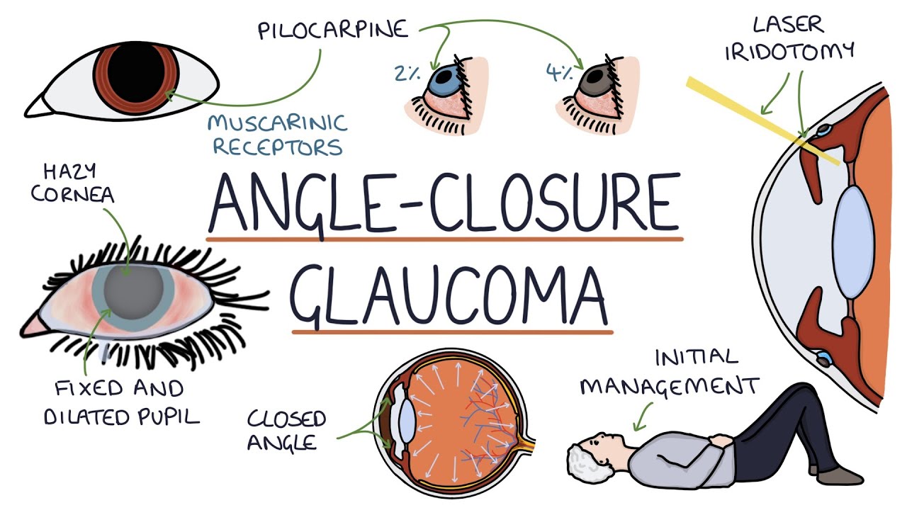
    -The two main types of glaucoma are open-angle and closed-angle glaucoma. The video focuses on open-angle glaucoma, where the resistance to the outflow of aqueous humor through the trabecular meshwork increases gradually.

  • How does aqueous humor flow through the eye, and how is it related to intraocular pressure?

    -Aqueous humor is produced by the ciliary body, flows from the posterior chamber to the anterior chamber, and drains through the trabecular meshwork into the canal of Schlemm. Resistance to this outflow creates intraocular pressure; if the resistance increases, the IOP rises.

  • What is the normal range of intraocular pressure, and what happens when this pressure increases in open-angle glaucoma?

    -The normal intraocular pressure is between 10 to 21 mmHg. In open-angle glaucoma, increased resistance in the trabecular meshwork leads to a gradual rise in pressure, causing damage to the optic nerve over time.

  • What is optic disc cupping, and how is it associated with glaucoma?

    -Optic disc cupping occurs when raised intraocular pressure causes the optic cup (a small indent in the optic disc) to become larger and deeper. An optic cup greater than 0.5 the size of the optic disc is considered abnormal and may indicate glaucoma.

  • What are the key risk factors for developing open-angle glaucoma?

    -Risk factors for open-angle glaucoma include increasing age, family history of glaucoma, black ethnic origin, and myopia (nearsightedness).

  • How does open-angle glaucoma typically present, and what are the common symptoms?

    -Open-angle glaucoma often presents asymptomatically, with a gradual loss of peripheral vision (tunnel vision), fluctuating pain, headaches, blurred vision, and halos around lights, especially at night.

  • What are the methods used to measure intraocular pressure, and how do they work?

    -Intraocular pressure is measured using non-contact tonometry, which estimates IOP by shooting a puff of air at the cornea, and Goldmann applanation tonometry, the gold standard, which applies pressure to the cornea to get an accurate measurement.

  • How is open-angle glaucoma diagnosed?

    -Diagnosis involves measuring intraocular pressure with Goldmann applanation tonometry, assessing optic disc cupping through fundoscopy, and checking for peripheral vision loss using visual field tests.

  • What are the treatment options for managing open-angle glaucoma?

    -Treatment aims to reduce intraocular pressure. First-line treatment includes prostaglandin analogs like latanoprost, which increase uveoscleral outflow. Other options include beta blockers, carbonic anhydrase inhibitors, sympathomimetics, and surgical procedures like trabeculectomy if eye drops are ineffective.

Outlines

00:00

๐Ÿ‘๏ธ Introduction to Open-Angle Glaucoma

Tom from Zero2Finals introduces open-angle glaucoma, explaining that it's caused by damage to the optic nerve due to a rise in intraocular pressure. The blockage of aqueous humor within the eye increases pressure, leading to glaucoma. He briefly outlines the types of glaucomaโ€”open and closed angleโ€”before diving into the basic anatomy and physiology of the eye, focusing on the aqueous humor's production and flow. The trabecular meshwork, the resistance it creates, and its role in regulating intraocular pressure are also explained.

05:01

๐Ÿง  Pathophysiology and Effects of Open-Angle Glaucoma

Tom explains that open-angle glaucoma develops gradually due to increased resistance in the trabecular meshwork, resulting in a slow buildup of intraocular pressure. This chronic pressure leads to cupping of the optic disc, where the optic cup expands due to pressure, indicating glaucoma if it exceeds half the size of the optic disc. Risk factors include age, family history, black ethnic origin, and myopia. He notes that early stages of the disease are often asymptomatic and detected during routine eye exams, with peripheral vision being affected first.

10:02

๐Ÿ‘“ Symptoms and Diagnosis of Open-Angle Glaucoma

Tom describes the symptoms of open-angle glaucoma, which include gradual peripheral vision loss, tunnel vision, fluctuating pain, headaches, and halos around lights, especially at night. He explains two methods to measure intraocular pressure: non-contact tonometry (using a puff of air) and Goldman applanation tonometry (the gold standard). These methods help diagnose glaucoma, alongside fundoscopy (to check for optic disc cupping) and visual field assessments (to detect peripheral vision loss).

๐Ÿ’Š Treatment and Management of Open-Angle Glaucoma

The goal of glaucoma treatment is to reduce intraocular pressure, especially when it reaches 24 mmHg or above. Prostaglandin analogs like latanoprost, which increase uveoscleral outflow, are the first-line treatment, though side effects include eyelash growth and iris pigmentation. Other treatments include topical beta blockers, carbonic anhydrase inhibitors, and sympathomimetics, all aiming to reduce aqueous humor production or increase its drainage. In severe cases, a trabeculectomy surgery may be performed to create a new drainage channel for the aqueous humor.

๐Ÿ“ข Conclusion and Resources

Tom concludes the video by encouraging viewers to explore more resources on Zero2Finals.com, including detailed notes, illustrations, questions, books, and flashcards. He also mentions his personal YouTube channel where he shares tips on learning medicine. He encourages viewers to subscribe and follow the channel for more content.

Mindmap

Keywords

๐Ÿ’กOpen-angle glaucoma

Open-angle glaucoma is a type of glaucoma where the drainage angle for the aqueous humor remains open, but the trabecular meshwork becomes gradually less efficient, leading to increased intraocular pressure. This slow buildup of pressure can cause optic nerve damage, often without noticeable symptoms in the early stages. The video focuses on this type of glaucoma, explaining its anatomy, causes, and treatment options.

๐Ÿ’กIntraocular pressure

Intraocular pressure (IOP) refers to the fluid pressure inside the eye. In the context of glaucoma, increased IOP is a key factor that causes optic nerve damage. The video details how a blockage in the trabecular meshwork can lead to higher IOP, causing glaucoma. A normal IOP range is 10-21 mmHg, and managing this pressure is essential to treating glaucoma.

๐Ÿ’กAqueous humor

Aqueous humor is the clear fluid produced by the ciliary body that fills the anterior and posterior chambers of the eye. It supplies nutrients to the cornea and lens and helps maintain intraocular pressure. In glaucoma, its impaired drainage through the trabecular meshwork leads to increased pressure. The video explains how this fluid flows through the eye and the consequences of its disrupted outflow.

๐Ÿ’กTrabecular meshwork

The trabecular meshwork is a spongy tissue located at the drainage angle between the cornea and the iris, responsible for regulating the outflow of aqueous humor. In open-angle glaucoma, this meshwork develops increased resistance, which prevents the fluid from exiting the eye properly, leading to elevated intraocular pressure. The video discusses this as the main site of dysfunction in open-angle glaucoma.

๐Ÿ’กOptic nerve

The optic nerve transmits visual information from the eye to the brain. In glaucoma, increased intraocular pressure can damage the optic nerve fibers, leading to vision loss. The video highlights how the optic nerve is affected in glaucoma, specifically focusing on the cupping of the optic disc as a result of prolonged elevated pressure.

๐Ÿ’กCupping of the optic disc

Cupping of the optic disc refers to the enlargement of the optic cup (a depression in the optic disc) due to increased intraocular pressure. When the optic cup becomes more than 0.5 times the size of the optic disc, it is considered abnormal and may indicate glaucoma. The video describes this as a key diagnostic feature of optic nerve damage in glaucoma.

๐Ÿ’กNon-contact tonometry

Non-contact tonometry is a method used to measure intraocular pressure by shooting a puff of air at the cornea and measuring its response. It is commonly used in optometry clinics for general screening, though it is less accurate than other methods. The video explains how this technique is used to detect potential cases of glaucoma.

๐Ÿ’กGoldman applanation tonometry

Goldman applanation tonometry is considered the gold standard for measuring intraocular pressure. It involves direct contact with the cornea using a device that applies pressure to get an accurate reading. The video contrasts this method with non-contact tonometry, noting its higher accuracy in diagnosing glaucoma.

๐Ÿ’กProstaglandin analogs

Prostaglandin analogs, such as latanoprost, are first-line treatments for open-angle glaucoma. These eye drops work by increasing the outflow of aqueous humor through the uveoscleral pathway, lowering intraocular pressure. The video mentions this as the primary treatment option for reducing IOP and managing glaucoma.

๐Ÿ’กTrabeculectomy

Trabeculectomy is a surgical procedure used to treat glaucoma when eye drops fail to lower intraocular pressure. It involves creating a new channel for aqueous humor to drain from the anterior chamber to a space under the conjunctiva, forming a 'bleb' where the fluid is reabsorbed. The video discusses this surgery as a last resort for managing open-angle glaucoma.

Highlights

Introduction to open-angle glaucoma, explaining it as optic nerve damage caused by a rise in intraocular pressure.

Open-angle glaucoma is due to increased resistance in the trabecular meshwork, leading to slow pressure buildup in the eye.

The anatomy of the eye, including the roles of the anterior and posterior chambers, filled with aqueous humor, is essential to understanding glaucoma.

Aqueous humor is produced by the ciliary body and drains through the trabecular meshwork into the canal of Schlemm.

Intraocular pressure normally ranges between 10 to 21 mmHg, with higher pressure indicating potential glaucoma.

Cupping of the optic disc is a key sign of glaucoma, where the optic cup becomes larger due to increased pressure.

Risk factors for open-angle glaucoma include age, family history, Black ethnic origin, and myopia (nearsightedness).

Open-angle glaucoma is often asymptomatic in early stages, detected during routine eye exams by checking intraocular pressure.

Peripheral vision is affected first in glaucoma, leading to tunnel vision as the disease progresses.

Glaucoma can also present with symptoms like fluctuating pain, headaches, blurred vision, and halos around lights, especially at night.

Non-contact tonometry is a common, less accurate way to measure intraocular pressure by using a puff of air.

Goldman applanation tonometry is the gold standard for measuring intraocular pressure, involving direct contact with the cornea.

Management of glaucoma aims to reduce intraocular pressure, with treatment often starting at 24 mmHg or above.

Prostaglandin analogues like latanoprost are first-line treatments, increasing the uveoscleral outflow to reduce eye pressure.

Surgical options, such as trabeculectomy, create a new drainage channel for aqueous humor when eye drops are ineffective.

Transcripts

play00:02

[Applause]

play00:03

[Music]

play00:10

hi this is tom from zero2finals.com

play00:12

in this video i'm going to be going

play00:14

through open angle glaucoma

play00:17

and you can find written notes on this

play00:18

topic at zero to finals dot com slash

play00:21

open angle glaucoma or in the

play00:23

ophthalmology section

play00:25

of the zero to finals medicine book

play00:28

so let's jump straight in glaucoma

play00:30

refers to the

play00:31

optic nerve damage that's caused by a

play00:35

significant rise in

play00:36

intraocular pressure the intraocular

play00:39

pressure is essentially the pressure

play00:41

within the eye

play00:43

itself raised intraocular pressure is

play00:46

caused by a blockage

play00:48

in the aqueous humor trying to escape

play00:50

the eye

play00:52

and we'll talk more about the

play00:53

pathophysiology shortly

play00:56

there are two types of glaucoma open

play00:59

angle

play01:00

and closed angle and here we're going to

play01:02

be talking about

play01:03

open angle so let's talk about the basic

play01:07

anatomy and physiology because it's

play01:10

important to understand

play01:12

some basic anatomy and physiology of the

play01:13

eye and how the eye

play01:15

functions in order to understand the

play01:17

pathophysiology

play01:19

of glaucoma the vitreous

play01:22

chamber of the eye is filled with

play01:24

vitreous humor

play01:26

and this is the main bulk the main area

play01:28

of the eye

play01:31

the anterior chamber is between the

play01:33

cornea and the iris

play01:36

and the posterior chamber is between the

play01:38

lens and the iris

play01:40

these anterior and posterior chamber are

play01:43

filled with

play01:44

aqueous humor that supplies nutrients to

play01:46

the cornea and keeps it healthy

play01:49

the aqueous humor is produced by the

play01:51

ciliary body

play01:52

and then the aqueous humor flows around

play01:55

the iris

play01:56

from the posterior chamber into the

play01:58

anterior chamber

play02:00

where it drains through something called

play02:02

the trabecular

play02:03

meshwork at the angle between the cornea

play02:06

and the iris

play02:09

the aqueous humor flows from the ciliary

play02:12

body

play02:13

around the lens and under the iris

play02:16

through the anterior chamber through the

play02:19

trabecular meshwork and

play02:20

into something called the canal of

play02:22

schlem

play02:23

from the canal of slam it eventually

play02:25

enters the general circulation

play02:28

and is reabsorbed into the body the

play02:30

normal

play02:31

intraocular pressure is 10 to 21

play02:35

millimeters of mercury and this pressure

play02:38

is created by the resistance to flow

play02:40

through the trabecular meshwork so

play02:43

aqueous humor

play02:44

is trying to flow through the trabecular

play02:47

meshwork

play02:48

out of the eye and if there's more

play02:51

resistance in the trabecular meshwork

play02:53

the pressure will go up because there's

play02:56

more resistance

play02:57

of aqueous humor trying to escape the

play02:59

eye if there's less resistance

play03:01

the pressure will go down because the

play03:03

aqueous humor is able to flow more

play03:05

freely out of the eye

play03:08

so let's talk about the pathophysiology

play03:10

of open-angle glaucoma

play03:11

and in open-angle glaucoma there's a

play03:13

gradual

play03:14

increase in resistance through the

play03:16

trabecular meshwork

play03:18

and this makes it more difficult for

play03:19

aqueous humor to flow through the

play03:21

meshwork and

play03:22

exit the eye therefore the pressure

play03:25

slowly builds within the eye

play03:27

and this gives a slow and chronic onset

play03:29

of glaucoma so let's talk about the

play03:32

effects of the increased pressure in the

play03:34

eye

play03:35

and this causes cupping of the optic

play03:37

disc

play03:40

and remember in the center of a normal

play03:42

optic disc is something called the optic

play03:44

cup and this is a small indent in the

play03:47

optic disc

play03:48

and is usually less than half the size

play03:50

of the optic cup

play03:51

and when there's a raised intraocular

play03:53

pressure this

play03:54

indent becomes larger because the

play03:56

pressure in the eye puts pressure on

play03:58

that indent making it

play04:00

wider and deeper this is called cupping

play04:04

an optic cup greater than 0.5 the size

play04:08

of the optic disc

play04:09

is abnormal so if the optic cup is more

play04:12

than half the size

play04:13

of the optic disc this suggests

play04:16

cupping and could indicate glaucoma

play04:20

so what are the risk factors for open

play04:22

angle glaucoma or increasing

play04:24

age family history black ethnic origin

play04:28

and near-sightedness which is called

play04:30

myopia are all

play04:32

key risk factors to remember for your

play04:34

exams

play04:36

let's talk about the presentation of

play04:37

open angle glaucoma

play04:39

often the rise in intraocular pressure

play04:41

is asymptomatic for a long period of

play04:44

time

play04:45

it's usually diagnosed by routine

play04:47

screening when they attend an

play04:48

optometry appointment for an eye check

play04:51

so whenever you get your eyes checked at

play04:52

the optometrist

play04:54

they check the intraocular pressure to

play04:57

see whether there's any possibility

play04:58

you've developed glaucoma

play05:00

glaucoma tends to affect the peripheral

play05:02

vision first

play05:04

so there may be a gradual loss of

play05:06

peripheral vision

play05:07

as it closes in slowly and they

play05:09

experience something called tunnel

play05:11

vision

play05:12

it can also present with gradual onset

play05:14

of fluctuating pain

play05:16

headaches blurred vision and typically

play05:20

halos surrounding lights which are worse

play05:22

at night time

play05:23

so if a patient is losing their

play05:25

peripheral vision and at night time they

play05:27

see halos appearing around lights

play05:30

think about glaucoma

play05:33

so how can we measure intraocular

play05:35

pressure well there's two

play05:37

main ways we can measure the intraocular

play05:39

pressure the first is something called

play05:41

non-contact

play05:42

tonometry and this is commonly used with

play05:45

a machine

play05:46

to estimate the intraocular pressure

play05:49

when you go to an opticians

play05:50

and this involves shooting a puff of air

play05:52

at the cornea

play05:54

and measuring the corneal response to

play05:56

that puff of air

play05:57

if the cornea has a lot of pressure

play05:59

inside it from glaucoma

play06:01

it won't react very much when air is

play06:04

shot at it

play06:05

whereas if you've got a very soft cornea

play06:07

with a lack of

play06:09

intraocular pressure there'll be a large

play06:11

reaction to that puff of air

play06:13

this is a less accurate way of measuring

play06:15

the intraocular pressure

play06:16

but it gives a helpful estimate for

play06:18

general screening purposes

play06:20

the second way of measuring intraocular

play06:22

pressure is something called

play06:23

goldman applination tonometry

play06:27

this is the gold standard way of

play06:29

measuring the intraocular pressure

play06:31

this involves using a special device

play06:33

mounted on a slip lamp

play06:34

that actually makes contact with the

play06:36

cornea and applies different pressures

play06:39

to the front of the cornea to get an

play06:40

accurate measurement of the intraocular

play06:42

pressure

play06:43

so the machine actually presses down on

play06:46

the cornea

play06:47

to check how much resistance there is

play06:50

and indicate how much pressure there is

play06:52

within that cornea

play06:55

so how do we make a diagnosis of

play06:57

glaucoma well firstly

play06:59

using a goldman applination tonometry

play07:02

which can be used to check the

play07:03

intraocular pressure

play07:04

fundoscopy is used to assess

play07:08

the optic disc for cupping and also for

play07:11

optic nerve health

play07:14

visual field assessment is important to

play07:16

check for peripheral vision loss

play07:18

and all three of these will be useful

play07:21

for making the diagnosis

play07:24

so how do we manage open angle glaucoma

play07:27

management of glaucoma aims to reduce

play07:29

the intraocular pressure

play07:31

treatment is usually started at an

play07:33

intraocular pressure

play07:34

of 24 millimeters of mercury or above

play07:37

patients should be closely followed up

play07:39

to assess the response to treatment

play07:41

and treatment will be guided by an

play07:43

ophthalmologist

play07:44

the first line treatment is something

play07:46

called prostaglandin analogues

play07:48

used as eye drops and an example of this

play07:51

is

play07:52

latanoprost and these eye drops increase

play07:55

the uveo

play07:56

scleral outflow and the uv scleral

play08:00

outflow refers to the drainage of the

play08:02

aqueous humor from the anterior chamber

play08:05

into the anterior chamber angle

play08:08

rather than going through the

play08:09

traditional trabecular meshwork

play08:12

notable side effects from prostaglandin

play08:14

analog eye drops like litanoprost

play08:17

are eyelash growth eyelid pigmentation

play08:21

and iris pigmentation typically you get

play08:24

a browning of the iris

play08:27

the other options for treating open

play08:29

angle glaucoma are

play08:30

topical beta blockers like timolol

play08:34

which reduce the production of the

play08:35

aqueous humor

play08:38

carbonic anhydrase inhibitors like

play08:40

dorsolamide

play08:41

which reduce the production of the

play08:43

aqueous humor and

play08:45

sympanometics such as brimonidine

play08:49

which reduce the production of aqueous

play08:51

fluid and also

play08:52

increase the uv scleral outflow

play08:55

finally a surgical procedure called

play08:58

trabeculectomy may be used

play09:00

where the eye drops are ineffective this

play09:02

involves creating a new

play09:03

channel from the anterior chamber

play09:06

through the sclera

play09:08

to a location under the conjunctiva this

play09:11

causes a little bleb

play09:13

under the conjunctival where the aqueous

play09:14

humor drains

play09:16

and the aqueous humor is then reabsorbed

play09:18

from this bleb into the general

play09:19

circulation

play09:20

so it essentially creates a new way for

play09:23

aqueous humor to drain away from the eye

play09:27

thank you for watching this video if you

play09:30

liked the video

play09:31

left a comment or subscribe to the

play09:33

channel thank you so much it really

play09:35

helps

play09:37

zero to finals is not just a youtube

play09:39

channel there's also a website with

play09:41

detailed notes

play09:42

illustrations and questions an instagram

play09:45

account where new questions are posted

play09:47

every day to help you test your

play09:49

knowledge

play09:50

books flash cards and much more

play09:55

i also have a personal channel where i

play09:57

share my thoughts and tips on learning

play09:58

medicine

play09:59

and you can find links to everything in

play10:01

the description of this video

play10:03

see you next time

Rate This
โ˜…
โ˜…
โ˜…
โ˜…
โ˜…

5.0 / 5 (0 votes)

Related Tags
GlaucomaOphthalmologyEye HealthDiagnosisTreatmentOptometryVision LossIntraocular PressureProstaglandin DropsEye Surgery