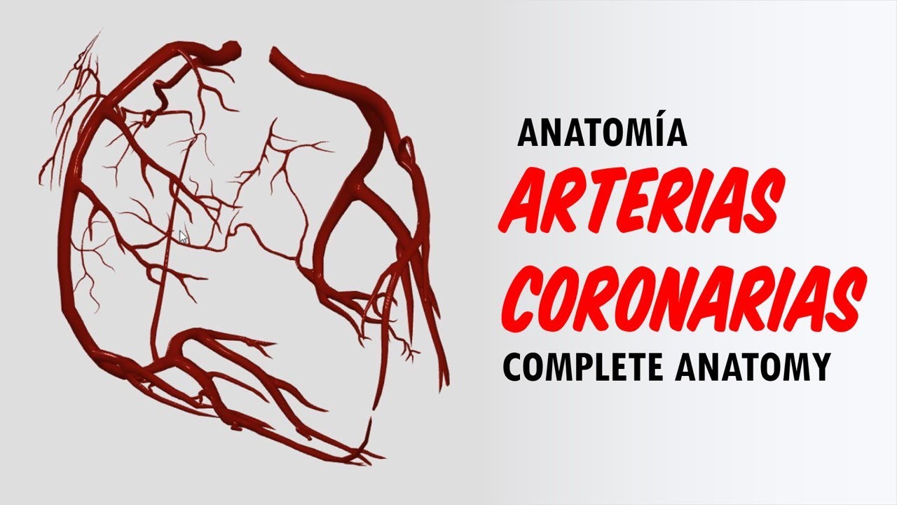
Coronary arteries Anatomy / Blood supply of Heart / Arterial supply of heart : Animation

Dr.G Bhanu Prakash Animated Medical Videos
29 Sept 201909:00

Summary

TLDRThis script delves into the arterial supply of the heart, detailing the coronary arteries' origins, courses, and branches. It explains the distribution of the right and left coronary arteries, their roles in supplying blood to different parts of the heart, and the clinical implications of their obstruction, such as angina pectoris and myocardial infarction. The script also covers the venous drainage of the heart, highlighting the coronary sinus and its tributaries, which are crucial for understanding cardiac function and related pathologies.

Takeaways

  • 🌟 The heart is primarily supplied by the right and left coronary arteries, which originate from the ascending aorta above the aortic valve.
  • πŸ” The right coronary artery (RCA) runs in the right atrioventricular groove and supplies the anterior surface of the pulmonary trunk, anterior ventricular branches, and atrial branches.
  • πŸ“ The left coronary artery (LCA) branches into the anterior interventricular artery and the circumflex artery, supplying various parts of the heart including the interventricular septum and left ventricle.
  • πŸ’Š Angina pectoris is a condition where narrowed coronary arteries reduce blood supply to the heart, causing chest pain during exertion that can radiate to the arm and shoulder.
  • πŸš‘ Myocardial infarction, or heart attack, occurs when a coronary artery is suddenly blocked, leading to ischemia and necrosis of the heart muscle.
  • ⚠️ The most common sites of coronary artery occlusion are the anterior interventricular artery, right coronary artery, and the circumflex branch of the left coronary artery.
  • 🩺 Clinical features of myocardial infarction include chest pain, nausea, vomiting, sweating, shortness of breath, and pain radiating to the arm and jaw.
  • πŸŒ€ The venous drainage of the heart involves the coronary sinus and various cardiac veins, which drain blood from the heart walls into the right atrium.
  • πŸ”„ The coronary sinus is the main vein of the heart, receiving blood from the great cardiac vein, middle cardiac vein, and small cardiac vein, among others.
  • πŸ”Ž The small cardiac vein accompanies the right ventricular artery and drains into the right end of the coronary sinus, while the oblique vein of the left atrium drains into the coronary sinus from the posterior surface of the left atrium.
  • πŸ”— The anterior cardiac veins and venae cordis minimae are small veins that drain blood from the right ventricle and all four chambers of the heart, respectively.

Q & A

  • What are the two main arteries that supply blood to the heart?

    -The heart is mostly supplied by the right and left coronary arteries, which arise from the ascending aorta.

  • Where do the coronary arteries originate from?

    -The right coronary artery originates from the anterior aortic sinus, and the left coronary artery originates from the left posterior aortic sinus, both located immediately above the aortic valve.

  • What is the anatomical feature that the coronary arteries lie within?

    -The coronary arteries and their branches run on the surface of the heart, lying within the subpericardial fibrofatty tissue.

  • How does the right coronary artery course after arising from the ascending aorta?

    -The right coronary artery first runs forwards between the pulmonary trunk and the right auricle, then descends vertically into the right atrioventricular groove, turns posteriorly at the inferior border of the heart, and terminates by anastomosing with the left coronary artery.

  • What are the main branches of the right coronary artery and what do they supply?

    -The right coronary artery supplies the anterior surface of the pulmonary conus, anterior ventricular branches, marginal branch, atrial branches (including the artery of the sinoatrial node in 60% of cases), and posterior ventricular branches, including the posterior interventricular artery.

  • What is the course of the left coronary artery after it arises from the ascending aorta?

    -The left coronary artery runs forwards and to the left between the pulmonary trunk and the left auricle, then divides into the anterior interventricular artery and the circumflex artery, which run in their respective grooves to the apex of the heart.

  • What is the function of the anterior interventricular artery, also known as the left anterior descending artery?

    -The anterior interventricular artery supplies the interventricular septum, the greater part of the left ventricle, and part of the right ventricle, as well as a part of the left bundle branch.

  • What clinical condition is associated with narrowed coronary arteries and how is it manifested?

    -Angina pectoris is associated with narrowed coronary arteries, causing moderate to severe pain in the region of the left precordium that may last up to 20 minutes, often referred to the left shoulder and medial side of the arm and forearm.

  • What is a myocardial infarction and what are its clinical features?

    -A myocardial infarction is a blockage of one of the larger branches of a coronary artery, leading to myocardial ischemia and necrosis. Clinical features include chest pain lasting longer than 30 minutes, nausea, vomiting, sweating, shortness of breath, and pain radiating to the arm, forearm, and hand.

  • Where do the three most common sites of coronary artery occlusion occur and what percentage do they represent?

    -The three most common sites of occlusion are the anterior interventricular artery (40-50%), the right coronary artery (30-40%), and the circumflex branch of the left coronary artery (15-20%).

  • How is the venous blood from the heart drained and what is the principal vein involved?

    -Venous blood from the heart is drained into the right atrium, primarily through the coronary sinus, which is the largest vein of the heart and receives blood from various tributaries including the great cardiac vein, middle cardiac vein, and small cardiac vein.

Outlines

00:00

πŸ’“ Arterial Supply and Branches of the Heart

The heart's arterial supply primarily comes from the right and left coronary arteries, which originate from the ascending aorta. The right coronary artery (RCA) supplies the anterior surface of the pulmonary conus, right ventricle, atria, and part of the interventricular septum. It also gives rise to the posterior interventricular artery. The left coronary artery (LCA) branches into the anterior interventricular artery (also known as the left anterior descending artery) and the circumflex artery. The anterior interventricular artery supplies the interventricular septum, the left ventricle, and part of the right ventricle, while the circumflex artery supplies the left ventricle's left margin and part of the left atrium. Clinically, narrowing of these arteries can lead to angina pectoris, characterized by chest pain during exertion that is relieved by rest.

05:01

πŸš‘ Myocardial Infarction and Venous Drainage

Myocardial infarction, commonly known as a heart attack, occurs when a coronary artery is suddenly blocked, leading to ischemia and subsequent necrosis of the heart muscle. This condition presents with symptoms such as prolonged chest pain, nausea, vomiting, sweating, shortness of breath, and pain radiating to the arm and jaw. The most common sites of blockage are the anterior interventricular artery (40-50%), the right coronary artery (30-40%), and the circumflex branch of the left coronary artery (15-20%). The heart's venous drainage system includes the coronary sinus and various cardiac veins that collect blood from the heart walls and return it to the right atrium. The coronary sinus is the largest vein and receives blood from the great cardiac vein, middle cardiac vein, small cardiac vein, and others, playing a crucial role in the heart's circulatory system.

Mindmap

Keywords

πŸ’‘Coronary Arteries

Coronary arteries are the blood vessels that supply oxygen-rich blood to the heart muscle. They are crucial for the heart's function and health. In the video's context, the right and left coronary arteries are described as arising from the aorta and branching out to supply different regions of the heart. The right coronary artery is mentioned as supplying the anterior surface of the pulmonary trunk and the right ventricle, while the left coronary artery divides into the anterior interventricular artery and the circumflex artery, supplying various parts of the heart.

πŸ’‘Subepicardial Fibrofatty Tissue

Subepicardial fibrofatty tissue is the layer of fat and connective tissue that lies beneath the pericardium, the sac surrounding the heart. This tissue is important for the structural support of the heart and provides a pathway for the coronary arteries to run on the surface of the heart. The script mentions that the coronary arteries and their branches lie within this tissue, emphasizing its role in the distribution of blood to the heart muscle.

πŸ’‘Right Coronary Artery (RCA)

The right coronary artery is one of the two main coronary arteries. It originates from the anterior aortic sinus and has a significant role in supplying blood to the right ventricle, the sinoatrial node, and parts of the left ventricle in some cases. The script describes its course from the aorta, its descent into the right atrioventricular groove, and its eventual anastomosis with the left coronary artery.

πŸ’‘Left Coronary Artery (LCA)

The left coronary artery is the other main coronary artery, arising from the left posterior aortic sinus. It is responsible for supplying blood to a large portion of the heart, including the interventricular septum and the left ventricle. The script details its path as it runs forward and to the left, dividing into the anterior interventricular artery and the circumflex artery.

πŸ’‘Anterior Interventricular Artery

Also known as the left anterior descending artery, this is a major branch of the left coronary artery. It runs down the anterior interventricular groove to the apex of the heart, supplying blood to the interventricular septum and parts of both ventricles. The script highlights its importance in the blood supply to the heart, making it a critical artery for cardiac function.

πŸ’‘Circumflex Artery

The circumflex artery is a branch of the left coronary artery that winds around the left margin of the heart. It supplies blood to the left margin of the left ventricle and is involved in the drainage of the pulmonary conus. The script describes its path and its termination at the posterior interventricular groove, where it anastomoses with the right coronary artery.

πŸ’‘Angina Pectoris

Angina pectoris is a medical condition characterized by chest pain due to insufficient blood supply to the heart muscle, typically occurring during exertion. The script explains that when coronary arteries are narrowed, the reduced blood supply can cause moderate to severe pain in the left precordial region, which can radiate to the left shoulder and arm.

πŸ’‘Myocardial Infarction

Myocardial infarction, commonly known as a heart attack, occurs when blood flow to a part of the heart is blocked, leading to ischemia and eventually necrosis of the heart muscle. The script describes the clinical features of a myocardial infarction, including chest pain, nausea, vomiting, sweating, and shortness of breath, and notes that it often results from the sudden blockage of a major coronary artery branch.

πŸ’‘Coronary Artery Occlusion

Coronary artery occlusion refers to the blockage of a coronary artery, which can lead to a heart attack. The script identifies the three most common sites of occlusion: the anterior interventricular artery, the right coronary artery, and the circumflex branch of the left coronary artery, each with a different percentage likelihood of being the site of blockage.

πŸ’‘Coronary Sinus

The coronary sinus is the principal vein of the heart, responsible for collecting most of the venous blood from the heart's walls and draining it into the right atrium. The script describes its location in the posterior part of the atrial ventricular groove and mentions its development from the left horn of the sinus venosus.

πŸ’‘Venous Drainage

Venous drainage refers to the process by which blood is returned to the heart from the body's tissues. In the context of the heart, the script discusses the various veins, such as the great cardiac vein, middle cardiac vein, and small cardiac vein, that contribute to the venous drainage by emptying into the coronary sinus.

Highlights

The heart is mostly supplied by the two coronary arteries which arise from the ascending aorta.

Coronary arteries and their branches run on the surface of the heart within the subpericardial fibrofatty tissue.

Anatomically, coronary arteries are not arteries but functionally behave like end arteries.

The right coronary artery arises from the anterior aortic sinus and runs between the pulmonary trunk and right atrium before descending into the right atrioventricular groove.

The right coronary artery supplies the anterior surface of the pulmonary conus, anterior ventricular branches, marginal branch, atrial branches, and posterior ventricular branches.

The left coronary artery arises from the left posterior aortic sinus and divides into the anterior interventricular artery and circumflex artery.

The anterior interventricular artery supplies the interventricular septum, greater part of the left ventricle, and part of the right ventricle.

The circumflex artery winds around the left margin of the heart and supplies the left marginal artery and diagonal artery.

Atrial branches of the coronary arteries supply the left atrium.

Narrowed coronary arteries can lead to reduced blood supply to cardiac muscles, causing angina pectoris during exertion.

Myocardial infarction occurs when a coronary artery is suddenly blocked, leading to ischemia and necrosis of the heart muscle.

The clinical features of myocardial infarction include chest pain, nausea, vomiting, sweating, shortness of breath, and pain radiating to the arm and jaw.

The three most common sites of coronary artery occlusion are the anterior interventricular artery, right coronary artery, and circumflex branch of the left coronary artery.

Venous blood from the heart is drained into the right atrium by the coronary sinus and other veins.

The coronary sinus is the principal vein of the heart, receiving blood from the walls of the heart.

The great cardiac vein, middle cardiac vein, and small cardiac vein are major tributaries of the coronary sinus.

The oblique vein of the left atrium and right marginal vein also contribute to the venous drainage of the heart.

Anterior cardiac veins and venae cordis minimae are small veins in the walls of the heart chambers that drain into the right atrium.

Transcripts

play00:00

[Music]

play00:05

arterial supply of the heart the heart

play00:09

is mostly supplied by the two coronary

play00:11

arteries which arise from the ascending

play00:13

aorta immediately above the eye or tick

play00:16

valve the coronary arteries and their

play00:18

branches run on the surface of the heart

play00:21

lying within the sub pericardial

play00:23

fibrofatty tissue anatomically coronary

play00:27

arteries are not and arteries but

play00:29

functionally they behave like end

play00:31

arteries right coronary artery origin

play00:35

the right coronary artery arises from

play00:38

the anterior attic sinus of the

play00:40

ascending aorta immediately above the I

play00:42

Arctic valve course after arising from

play00:46

the ascending aorta the right coronary

play00:48

artery first runs forwards between the

play00:51

pulmonary trunk and the right oracle and

play00:54

then it descends almost vertically into

play00:57

the right atrioventricular groove the

play01:00

right anterior coronary sulcus up to the

play01:03

junction of the right and inferior

play01:05

borders of the heart

play01:07

at the inferior border of the heart it

play01:09

turns posteriorly and runs in the

play01:12

posterior atrioventricular groove up to

play01:15

the posterior interventricular groove

play01:17

and terminates by anastomosing with the

play01:20

left coronary artery

play01:22

branches and distribution right CONUS

play01:26

artery it supplies the anterior surface

play01:28

of the pulmonary CONUS anterior

play01:31

ventricular branches they are two or

play01:35

three and supply the anterior surface of

play01:37

the right ventricle

play01:38

the marginal branch is the largest and

play01:41

runs along the lower margin of the

play01:43

sternal costal surface to reach the apex

play01:46

atrial branches they supply the atria

play01:49

one of the atrial branches the artery of

play01:52

the sinoatrial node supplies the SA node

play01:55

in 60% of cases in 40 percent of

play01:58

individuals it arises from the left

play02:00

coronary artery

play02:02

posterior ventricular branches they are

play02:05

usually to and supply the diaphragmatic

play02:07

surface of the right and left ventricles

play02:11

posterior interventricular artery it

play02:14

runs in the posterior interventricular

play02:16

groove up to the apex it supplies the

play02:20

posterior part of the interventricular

play02:22

septum AV node in 60% cases and right

play02:26

and left ventricles

play02:29

left coronary artery origin the left

play02:33

coronary artery arises from the left

play02:35

posterior I Arctic sinus of the

play02:37

ascending aorta immediately above the I

play02:40

Arctic valve course after arising from

play02:44

the ascending aorta the left coronary

play02:46

artery runs forwards and to the left

play02:49

between the pulmonary trunk and the left

play02:52

auricle it then divides into an anterior

play02:55

interventricular artery also known as

play02:58

left anterior descending artery and runs

play03:01

downwards in the anterior

play03:03

interventricular groove to the apex of

play03:05

the heart it then passes posteriorly

play03:08

around the apex of the heart to enter

play03:11

the posterior interventricular groove to

play03:14

terminate by anastomosing with the

play03:17

posterior interventricular artery a

play03:19

branch of the right coronary artery the

play03:22

circumflex artery winds around the left

play03:25

margin of the heart and continues in the

play03:27

left posterior coronary sulcus up to the

play03:30

posterior interventricular groove where

play03:33

it terminates by anastomosing with the

play03:36

right coronary artery

play03:39

branches and distribution anterior

play03:42

interventricular artery also known as

play03:44

left anterior descending artery it

play03:46

supplies and here you're part of

play03:49

interventricular septum greater part of

play03:52

the left ventricle and part of the right

play03:54

ventricle and a part of left bundle

play03:57

branch circumflex artery it gives a left

play04:01

marginal artery that supplies the left

play04:04

margin of the left ventricle up to the

play04:06

apex of the heart diagonal artery it may

play04:10

arise directly from the trunk of the

play04:13

left coronary artery CONUS artery it

play04:16

supplies the pulmonary CONUS atrial

play04:20

branches they supply the left atrium

play04:23

clinical correlation angina pectoris

play04:27

if the coronary arteries are narrowed

play04:29

the blood supply to the cardiac muscles

play04:32

is reduced as a result on exertion the

play04:35

patient feels moderately severe pain in

play04:38

the region of left procore diem that may

play04:41

last as long as 20 minutes the pain is

play04:44

often referred to the left shoulder and

play04:46

medial side of the arm and forearm in

play04:49

angina pectoris pain occurs on exertion

play04:53

and relieved by rest this is because the

play04:56

coronary arteries are so narrowed that

play04:58

ischemia of cardiac muscle occurs only

play05:01

on exertion

play05:03

myocardial infarction a sudden block of

play05:06

one of the larger branches of either

play05:08

coronary artery usually leads to

play05:10

myocardial ischemia followed by the

play05:13

myocardial necrosis which is myocardial

play05:15

infarction the part of heart suffering

play05:19

from myocardial infarction stops

play05:21

functioning and often causes death this

play05:24

condition is termed heart attack or

play05:26

coronary attack the clinical features of

play05:29

myocardial infarction are as follows a

play05:32

sensation of pressure or sinking and

play05:35

pain in the chest that lasts longer than

play05:37

30 minutes nausea or vomiting sweating

play05:41

shortness of breath and tachycardia pain

play05:46

radiates to the medial side of the arm

play05:48

forearm and hand sometimes it may be

play05:51

referred to jaw or neck sites of

play05:55

coronary artery occlusion the three most

play05:58

common sites of the coronary artery

play06:00

occlusion are as under anterior

play06:03

interventricular artery or left anterior

play06:06

descending artery is 40 to 50 percent

play06:09

right coronary artery is 30 to 40

play06:12

percent circumflex branch of the left

play06:15

coronary artery is 15 to 20 percent an

play06:18

important point to be noted here is that

play06:21

myocardial infarction mostly occurs at

play06:23

breast where as angina occurs on

play06:26

exertion anterior interventricular

play06:28

artery or left anterior descending

play06:31

artery is most commonly blocked venous

play06:34

drainage of the heart venous blood from

play06:37

the heart is drained into right atrium

play06:39

by the following coronary sinus anterior

play06:43

cardiac veins vena cordis mini-me also

play06:47

known as the Bayesian veins coronary

play06:50

sinus it is the principal vein of the

play06:52

heart most of the venous blood from the

play06:55

walls of the heart is drained into the

play06:57

right atrium through coronary sinus the

play07:00

coronary sinus is the largest vein of

play07:03

the heart and lies in the posterior part

play07:05

of the atrial ventricular groove it

play07:08

develops from the left horn of the sinus

play07:10

venous and a part of the left common

play07:12

Cardinal vein tributaries the

play07:16

coronary sinus receives the following

play07:17

tributaries great cardiac vein it

play07:21

accompanies anterior interventricular

play07:23

and circumflex arteries to join the left

play07:26

end of the coronary sinus middle cardiac

play07:29

vein it accompanies the posterior

play07:32

interventricular artery to join the

play07:34

coronary sinus near its termination

play07:37

small cardiac vein it accompanies the

play07:40

right ventricular artery in the right

play07:42

posterior coronary sulcus and the right

play07:45

end of the coronary sinus posterior Wain

play07:48

of the left ventricle it runs on the

play07:51

diaphragmatic surface of the left

play07:53

ventricle and joins the sinus to the

play07:55

middle cardiac vein oblique vein of left

play07:59

atrium also known as vein of Marshall it

play08:02

is a small wind which runs downwards on

play08:05

the posterior surface of the left atrium

play08:07

to enter the left end of the coronary

play08:10

sinus it develops from the left common

play08:12

Cardinal vein which is also known as a

play08:15

duct of Q veer right marginal vein it

play08:19

accompanies the marginal branch of the

play08:22

right coronary artery and joins the

play08:24

small cardiac vein or drains directly

play08:26

into the right atrium left marginal vein

play08:30

it accompanies the marginal branch of

play08:32

the left coronary artery and drains into

play08:35

the coronary sinus anterior cardiac

play08:38

veins these are a series of small veins

play08:41

which run parallel to each other across

play08:44

the surface of right ventricle to open

play08:46

into the right atrium vena cordis

play08:49

minimize also known as the Bayesian

play08:52

veins these are extremely small veins in

play08:55

the walls of all four chambers of the

play08:57

heart

Rate This
β˜…
β˜…
β˜…
β˜…
β˜…

5.0 / 5 (0 votes)

Related Tags
Coronary ArteriesHeart HealthCardiac SupplyArterial AnatomyAngina PectorisMyocardial InfarctionHeart AttackVenous DrainageCardiologyHealth EducationMedical Anatomy