🥇 PLEXO CERVICAL, Anatomía (Superficial y Profundo). Fácil, Rápido y Sencillo
Summary
TLDREn este video, Juan José Sánchez nos guía a través de la anatomía del cuello, centrando la discusión en la innvación cervical. Se detalla la formación del plexo cervical, compuesto por las ramas anteriores de los nervios cervicales desde C1 hasta C4, y se divide en el plexo cervical superficial y profundo. El primero se encarga de la innvación cutánea del cuello, formado por nervios como el nervio occipital menor, el nervio auricular mayor, el nervio cervical transverso y el nervio supraclavicular. Por otro lado, el plexo cervical profundo es motor y proporciona innvación a los músculos del región anterolateral del cuello, incluyendo el músculo esternocleidomastoideo, trapézio y levador de la escápula. Además, se menciona la formación y función del nervio frénico, que innvación al diafragma y tiene una relación anatómica importante con el músculo escaleno anterior. Finalmente, se destaca la innvación de los músculos prevertebrales y los músculos infrahioideos, excluyendo el músculo tirohioideo, que es innervado directamente por las ramas anteriores de C1 y C2.
Takeaways
- 📚 El plexo cervical es una red de nervios formada por las ramas anteriores de los primeros cuatro nervios cervicales (C1 a C4).
- 🌐 El plexo cervical se divide en dos partes: el plexo cervical superficial y el plexo cervical profundo.
- 🔍 El plexo cervical superficial es responsable de la innervación cutánea del cuello, incluyendo la sensibilidad a la dolor, calor y frío.
- 💡 Los cuatro nervios que componen el plexo cervical superficial son el nervio occipital menor, el nervio auricular mayor, el nervio transverso cervical y el nervio supraclavicular.
- 🏋️ El plexo cervical profundo tiene una función motora, innervando los músculos del región anterolateral del cuello, incluyendo el músculo esternocleidomastoideo y el trapezio.
- 🔑 El plexo cervical profundo también da lugar a dos estructuras importantes: la bobina cervical y el nervio frénico.
- 🤔 La bobina cervical, formada por las uniones de C1 y C2 en la raíz superior y C2 y C3 en la raíz inferior, innerva los músculos infrahioideos, excepto el músculo tirohioideo.
- 🫁 El nervio frénico, compuesto por las ramas anteriores de C3, C4 y C5, tiene su principal función en la innervación del diafragma, aunque también proporciona sensibilidad a las serosas del tórax y abdomen.
- ⚙️ Además de la bobina cervical y el nervio frénico, el plexo cervical profundo emite ramas musculares específicas para otros músculos del cuello.
- 📌 Es importante destacar que la innervación del cuello no proviene de todas las ramas de los nervios C1 a C4, sino que puede ser una combinación de algunas de ellas.
- 🧲 La innervación cutánea del cuello por el plexo cervical superficial cubre áreas específicas, mientras que el plexo cervical profundo tiene una función motora y sensorial en otros tejidos.
Q & A
¿Qué es el plexo cervical y qué nervios forma?
-El plexo cervical es una red de nervios formada por la unión de las ramas anteriores de los primeros cuatro nervios cervicales, desde C1 hasta C4.
¿Cómo se divide el plexo cervical y cuál es la diferencia principal entre las dos divisiones?
-El plexo cervical se divide en un plexo superficial y un plexo profundo. La principal diferencia es que el plexo superficial es claramente sensible, proporcionando innervación cutánea al cuello, mientras que el plexo profundo es motor, proporcionando innervación a los músculos del cuello.
¿Cuáles son los cuatro nervios que componen el plexo cervical superficial?
-Los cuatro nervios que componen el plexo cervical superficial son el nervio occipital menor, el nervio auricular mayor, el nervio transverso cervical y el nervio supraclavicular.
¿Qué nervios componen el plexo cervical profundo y cuál es su principal función?
-El plexo cervical profundo está compuesto por dos grandes estructuras: la boucle cervical y el nervio frénico. Su principal función es proporcionar innervación motora a los músculos del cuello.
¿Qué nervio está formado por las ramas anteriores de los nervios C3, C4 y C5 y cuál es su función principal?
-El nervio frénico está formado por las ramas anteriores de los nervios C3, C4 y C5, y su función principal es innervar el diafragma.
¿Cuál es la relación anatómica del nervio frénico con el músculo anterior escaleno?
-El nervio frénico siempre está frente al músculo anterior escaleno mientras desciende.
¿Cómo se relaciona el nervio frénico con la pleura y el pericardio?
-El nervio frénico sensiblemente innerva la serosa del tórax y el abdomen, lo que incluye la pleura y el pericardio.
¿Qué músculos del cuello son innervados por el plexo cervical profundo a excepción de la boucle cervical y el nervio frénico?
-El plexo cervical profundo también proporciona ramas musculares a los músculos esternocleidomastoideo, trapezius, escaleno y los cuatro músculos prevertebrales.
¿Cómo se llama la estructura que forma la boucle cervical y qué músculos innerva?
-La boucle cervical se forma a partir de C1 a C3, incluyendo C2, y innerva todos los músculos infrahioideos excepto el músculo tirohioideo.
¿Cuál es la innervación del músculo tirohioideo y cómo se relaciona con el nervio hipogloso?
-El músculo tirohioideo es innervado por una rama directa de C1 y C2, no por el nervio hipogloso, a pesar de que antiguamente se pensó que lo era.
¿Qué nervios componen la primera rama del plexo cervical superficial y cuál es su función?
-La primera rama es el nervio occipital menor, que proporciona innervación sensorial al área lateral de la cabeza y a la parte craneal del oído.
¿Cómo se relacionan los nervios del plexo cervical superficial con el músculo esternocleidomastoideo y la vena yugular interna?
-Los nervios del plexo cervical superficial emergen detrás o profundamente a la vena yugular interna y detrás del músculo esternocleidomastoideo, volviéndose muy superficiales justo en el borde posterior del esternocleidomastoideo.
Outlines

このセクションは有料ユーザー限定です。 アクセスするには、アップグレードをお願いします。
今すぐアップグレードMindmap

このセクションは有料ユーザー限定です。 アクセスするには、アップグレードをお願いします。
今すぐアップグレードKeywords

このセクションは有料ユーザー限定です。 アクセスするには、アップグレードをお願いします。
今すぐアップグレードHighlights

このセクションは有料ユーザー限定です。 アクセスするには、アップグレードをお願いします。
今すぐアップグレードTranscripts

このセクションは有料ユーザー限定です。 アクセスするには、アップグレードをお願いします。
今すぐアップグレード関連動画をさらに表示

🥇 Anatomía del ESÓFAGO, Fácil y Rápida

🥇 HUESO HIOIDES, ANATOMÍA. ¡Explicación Sencilla!

🥇 HUESO OCCIPITAL, Anatomía. Fácil, Rápido y Sencillo
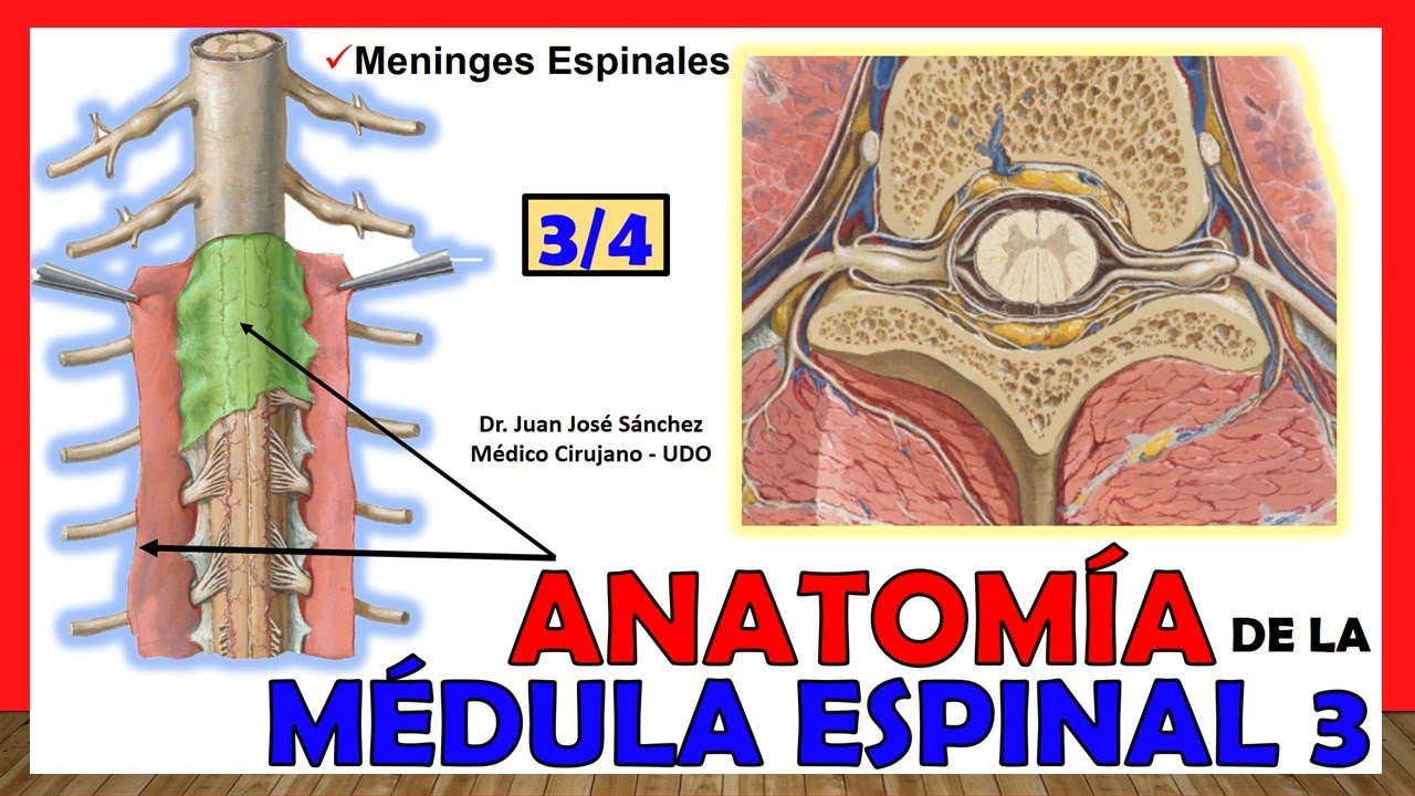
🥇 MÉDULA ESPINAL 3/4 - (Meninges Espinales) -Anatomía. ¡Explicación fácil!

🥇 Articulación TEMPOROMANDIBULAR. Fácil, Rápida y Sencilla

🥇 ARACNOIDES 2/2 - Cisternas Subaracnoideas. ¡Explicación Sencilla!
5.0 / 5 (0 votes)
