HSCI 4950- Histology Basement membrane
Summary
TLDRThis educational video script delves into the structure and function of the basement membrane, a critical component beneath epithelial cells. It discusses the composition of the basal lamina and reticular lamina, highlighting the roles of fibroblasts and collagen. The script explains how the basement membrane, visible under an electron microscope, serves as a support and regulatory layer for epithelial cells, controlling macromolecule access and facilitating nerve reinnervation. It also mentions the presence of growth factor binding sites, crucial for tissue development.
Takeaways
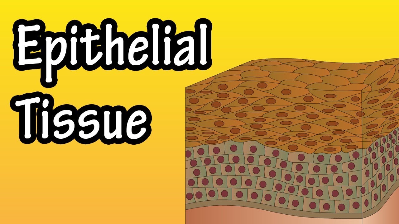
- 🔬 Epithelial cells have various structures on their apical surface, including microvilli and cilia, which aid in their function.
- 🔗 Cell junctions such as desmosomes, gap junctions, and tight junctions are found on the lateral surfaces of epithelial cells, facilitating cell-cell connections.
- 🏗️ The basement membrane is located at the bottom of epithelial cells and is composed of the basal lamina and the reticular lamina.
- 👨🔬 Fibroblasts secrete the connective tissue fibers, primarily collagen type 3, which are found beneath the epithelial cells.
- 💡 The basal lamina is visible only under an electron microscope and consists of a lamina lucida and a lamina densa, containing collagen type 4 and intermediate filaments.
- 🌱 The basal lamina is secreted by epithelial cells and forms a connection to the underlying connective tissue.
- 🔍 The reticular lamina, also known as the connective tissue fibers, is secreted by fibroblasts and is part of the basement membrane.
- 📚 The basement membrane can be visualized using the periodic acid-Schiff (PAS) stain in a light microscope.
- 🛡️ The basement membrane serves two main functions: providing support to the epithelial cells and regulating the access of macromolecules to the cells.
- 📈 The basal lamina contains binding sites for growth factors, which are crucial for tissue development and directionality.
- 🤝 The basement membrane plays a role in cell-cell interactions, especially in nerve reinnervation after injury.
Q & A
What is the basement membrane and where is it located?
-The basement membrane is a layer of extracellular matrix found at the bottom of epithelial cells, serving as a connection between the epithelial cells and the underlying connective tissue.
What are the two main components of the basement membrane when viewed under an electron microscope?
-The two main components are the lamina lucida, which is not always present, and the lamina densa, which contains mostly collagen type 4 and an intermediate filament known as laminin.
What cells are responsible for secreting the fibers found in the connective tissue layer beneath the basement membrane?
-Fibroblasts are the cells that secrete the fibers, primarily collagen type 3 and other intermediate filaments, in the connective tissue layer.
What is the term used for the connective tissue fibers that are secreted by fibroblasts and are associated with the basement membrane?
-The connective tissue fibers are sometimes referred to as the reticular lamina.
What does the term 'lamina propria' refer to and what is its function?
-The term 'lamina propria' refers to the supportive layer beneath certain epithelia, and its function is to provide support to the epithelial cells.
How does the basement membrane contribute to the regulation of macromolecules accessing the epithelial cells?
-The basement membrane can control the access of macromolecules to the epithelial cells above, acting as a selective barrier.
What role do growth factors play in relation to the basal lamina during tissue development?
-Growth factors, which are important for tissue development and directional growth, often have binding sites on the basal lamina, guiding the development of epithelial tissue.
How is the basement membrane involved in nerve reinnervation after a nerve is severed?
-The basement membrane facilitates the process of nerve reinnervation by aiding the nerve fibers as they attempt to reconnect within the nerve sheath.
What staining technique is used to visualize the basement membrane in the light microscope?
-The periodic acid-Schiff (PAS) stain is used to visualize the basement membrane in the light microscope.
What is the significance of the basement membrane in terms of cell-cell interactions?
-The basement membrane plays a crucial role in cell-cell interactions, particularly in processes like nerve reinnervation, where it helps guide and support cellular reconnections.
Why is the lamina lucida not always visible in the basement membrane?
-The lamina lucida may not always be present due to variations in tissue types and the specific conditions under which the basement membrane is examined.
Outlines

Dieser Bereich ist nur für Premium-Benutzer verfügbar. Bitte führen Sie ein Upgrade durch, um auf diesen Abschnitt zuzugreifen.
Upgrade durchführenMindmap

Dieser Bereich ist nur für Premium-Benutzer verfügbar. Bitte führen Sie ein Upgrade durch, um auf diesen Abschnitt zuzugreifen.
Upgrade durchführenKeywords

Dieser Bereich ist nur für Premium-Benutzer verfügbar. Bitte führen Sie ein Upgrade durch, um auf diesen Abschnitt zuzugreifen.
Upgrade durchführenHighlights

Dieser Bereich ist nur für Premium-Benutzer verfügbar. Bitte führen Sie ein Upgrade durch, um auf diesen Abschnitt zuzugreifen.
Upgrade durchführenTranscripts

Dieser Bereich ist nur für Premium-Benutzer verfügbar. Bitte führen Sie ein Upgrade durch, um auf diesen Abschnitt zuzugreifen.
Upgrade durchführenWeitere ähnliche Videos ansehen

Epithelial Tissue Histology Explained for Beginners | Corporis

Tissues of Human Body | Animation | Simple Explanation

BIOLOGI Kelas 11 - Jaringan Hewan (Part 1) | GIA Academy

Intermediate filaments Part 2
Epithelial Tissue - What Is Epithelial Tissue - Functions Of Epithelial Tissue - Epithelial Cells
BAB 1 : MENJELAJAH SEL | Biologi Kelas 11 Kurikulum Merdeka
5.0 / 5 (0 votes)
