Central nervous system: Histology
Summary
TLDRThis script explores the central nervous system (CNS), highlighting its key components: the cerebrum, cerebellum, brainstem, and spinal cord. It delves into the roles of neurons and neuroglia, emphasizing their functions in the nervous system. The script contrasts the CNS's white and gray matter, explaining their composition and appearance. It also discusses the meninges, cerebrospinal fluid, and various cell types, including astrocytes and microglia, detailing their unique characteristics and roles. The video provides a comprehensive look at the CNS's structure and function, crucial for understanding neurological processes.
Takeaways
- 🧠 The central nervous system (CNS) comprises the cerebellum, cerebrum, brainstem, and spinal cord.
- 🌐 Neurons are the fundamental working units of the nervous system, while neuroglia or glial cells provide support and protection.
- 🧬 Central neuroglia includes astrocytes, oligodendrocytes, endothelial cells, and microglia, whereas peripheral neuroglia includes Schwann cells, satellite cells, and other organ-associated cells.
- 🔍 The CNS is macroscopically divided into white matter, rich in myelinated axons, and gray matter, primarily composed of neuron cell bodies and dendrites.
- 👀 In the spinal cord, white matter is peripheral, and gray matter is central, creating an H or butterfly shape in cross-section.
- 🛡️ The meninges are connective tissue layers that envelop the CNS, with the dura mater being the thickest and outermost layer.
- 💧 The brain's ventricles and the spinal cord's central canal are filled with cerebrospinal fluid (CSF), produced by ependymal cells.
- 🔬 Astrocytes are the most abundant glial cells in the CNS, with processes that help form the blood-brain barrier and produce glial fibrillary acidic protein (GFAP).
- 🔍 Microglia are the immune cells of the CNS, capable of moving and migrating to remove damaged tissue and participate in immune responses.
- 🧠 Pyramidal cells are prominent in the cerebral cortex, integrating sensory information and initiating motor responses, characterized by a large cell body and a single axon.
- ⚖️ The cerebellar cortex has three layers: the white matter, the granular layer, and the molecular layer, with Purkinje cells situated between the molecular and granular layers.
Q & A
What are the main components of the central nervous system (CNS)?
-The central nervous system (CNS) consists of the cerebellum, cerebrum, brainstem, and spinal cord.
What is the basic working unit of the nervous system?
-The neuron is the basic working unit of the nervous system.
What role do neuroglia or glial cells play in the nervous system?
-Neuroglia or glial cells are non-neuronal cells that support and protect the nervous system.
What are the differences between white matter and gray matter in the CNS?
-White matter is composed of lipid-rich myelin sheaths covering axons, while gray matter consists mostly of neuron cell bodies, dendrites, astrocytes, and microglia.
How is the spinal cord's arrangement of white and gray matter different from the brain?
-In the spinal cord, white matter is mainly in the periphery, and gray matter is closer to the center, forming an H or butterfly-shaped appearance in cross-section, which is the opposite arrangement found in the brain.
What is the function of the meninges in the CNS?
-The meninges are layers of connective tissue that cover and protect the CNS, with the dura mater being the thickest and outermost layer.
What is cerebrospinal fluid (CSF) and what is its function?
-Cerebrospinal fluid (CSF) is a clear fluid that fills the ventricles of the brain and the central canal of the spinal cord, providing cushioning and support to the CNS.
What are the characteristics of astrocytes and how do they contribute to the blood-brain barrier?
-Astrocytes are star-shaped glial cells with long processes that surround capillaries, helping to maintain the blood-brain barrier by inducing and maintaining tight junctions between endothelial cells.
How do oligodendrocytes contribute to the CNS?
-Oligodendrocytes are glial cells that wrap around axons to form myelin sheaths within the CNS, aiding in the speed of nerve impulse conduction.
What is the role of microglia in the CNS?
-Microglia are the immune cells of the CNS, responsible for finding and removing damaged tissue and participating in the CNS immune response.
What are the characteristics of pyramidal cells and their role in the cerebral cortex?
-Pyramidal cells are prominent neurons in the cerebral cortex with a large, pyramid-shaped cell body and a branching dendritic system. They integrate sensory information and initiate voluntary motor responses.
How does the cerebellum's cortex differ in structure from the cerebral cortex?
-The cerebellum's cortex has a distinct three-layered structure with the molecular layer, a single layer of Purkinje cells, and the granular layer, which is different from the six-layered structure of the cerebral cortex.
Outlines

Dieser Bereich ist nur für Premium-Benutzer verfügbar. Bitte führen Sie ein Upgrade durch, um auf diesen Abschnitt zuzugreifen.
Upgrade durchführenMindmap

Dieser Bereich ist nur für Premium-Benutzer verfügbar. Bitte führen Sie ein Upgrade durch, um auf diesen Abschnitt zuzugreifen.
Upgrade durchführenKeywords

Dieser Bereich ist nur für Premium-Benutzer verfügbar. Bitte führen Sie ein Upgrade durch, um auf diesen Abschnitt zuzugreifen.
Upgrade durchführenHighlights

Dieser Bereich ist nur für Premium-Benutzer verfügbar. Bitte führen Sie ein Upgrade durch, um auf diesen Abschnitt zuzugreifen.
Upgrade durchführenTranscripts

Dieser Bereich ist nur für Premium-Benutzer verfügbar. Bitte führen Sie ein Upgrade durch, um auf diesen Abschnitt zuzugreifen.
Upgrade durchführenWeitere ähnliche Videos ansehen

Anatomi Sistem Saraf: Saraf Pusat & Tepi | Neurologi
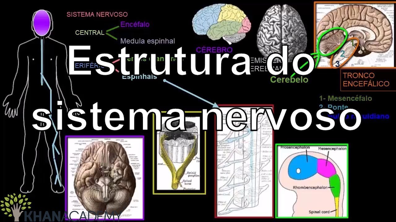
Estrutura do sistema nervoso | Saúde e medicina anatomia e fisiologia humana | Khan Academy

SISTEM KOORDINASI PADA MANUSIA - KELAS XI

Como funciona o Sistema Nervoso Central?
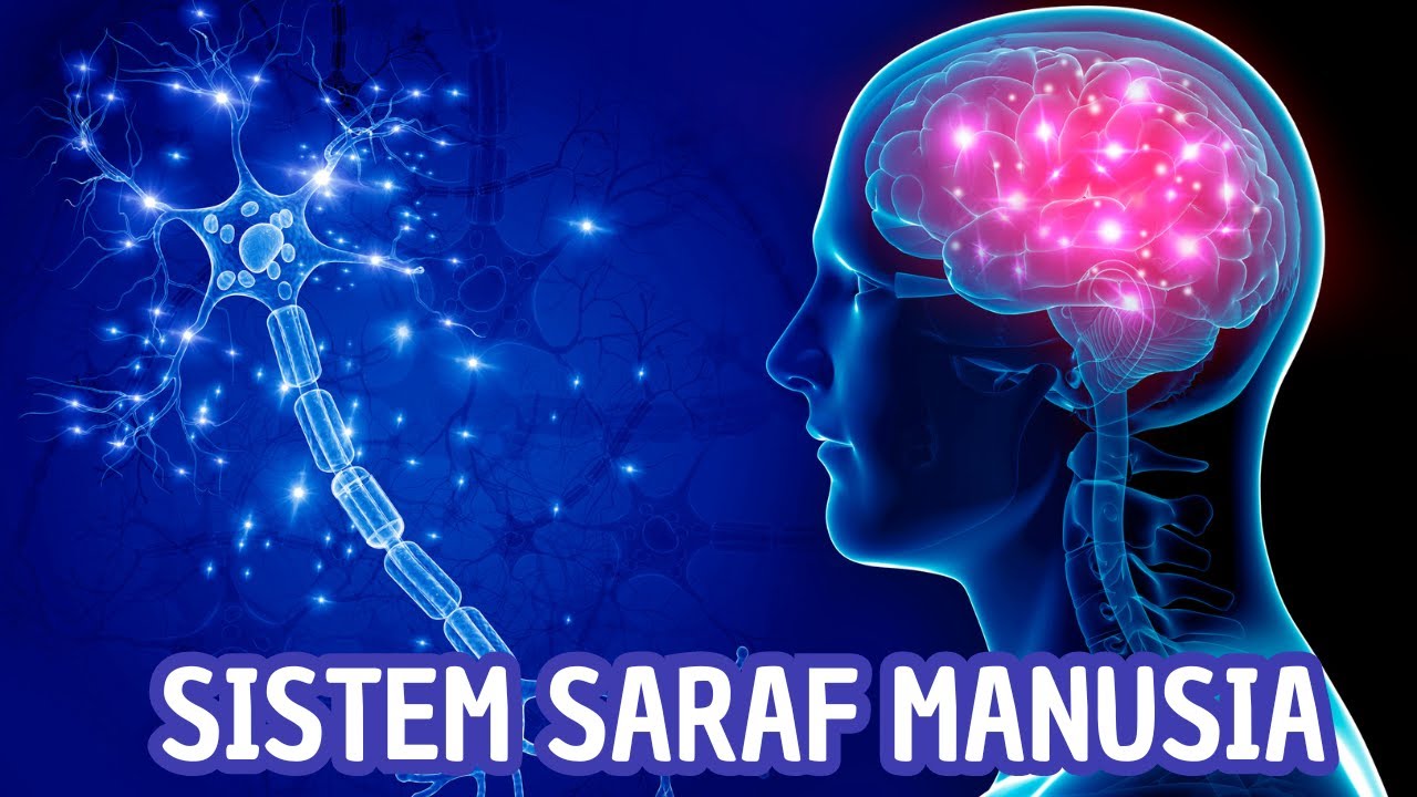
Sistem Saraf Manusia: Bagian-Bagian dan Fungsinya

Centrala nervsystemets uppbyggnad och funktion
5.0 / 5 (0 votes)
