What is DICOM | PACS & VNA Fundamentals
Summary
TLDRThis video delves into the DICOM standard for medical imaging, its integration with PACS (Picture Archiving Communication Systems), and the evolution towards VNA (Vendor Neutral Archive). It traces the history from X-ray film to digital radiography, explaining how DICOM files and protocols facilitate image transfer and storage. The video also discusses the shift from physical to digital annotations and the challenges of a VNA in handling diverse medical imaging data, including non-DICOM formats.
Takeaways
- 📚 The video discusses the DICOM standard used in medical imaging and its relation to PACS (Picture Archiving Communication Systems) and VNA (Vendor Neutral Archive).
- 🏥 Historically, X-rays were printed on films and processed in dark rooms, but now they are transferred digitally to radiologist's monitors in DICOM format.
- 🏅 Notable figures like William Rankin, Marie Curie, Pierre Curie, and Henri Becquerel contributed significantly to the field of radiation and medical imaging.
- 🔖 In traditional methods, X-ray films were organized with patient demographics in physical folders, following a hanging protocol for easy review by radiologists.
- 📈 The advent of digital technology has made the process of medical imaging completely digital, with images and patient data combined into DICOM files for easy transfer and storage.
- 🖼️ DICOM files are not just images; they include patient metadata, referred to as DICOM tags, which are crucial for organizing and retrieving medical images.
- 💾 PACS is a digital solution that has replaced the traditional film-based workflow in radiology departments, allowing for the storage, transfer, and viewing of medical images.
- 📊 VNA is an extension of PACS, aiming to be a single solution for storing, transmitting, and viewing all types of medical images across different departments and vendors.
- 🔍 Radiologists can now make digital annotations on images, which are stored within the PACS as part of the DICOM file, enhancing the review process.
- 🌐 The challenge for VNAs is to not only store but also view a multitude of DICOM and non-DICOM images, which may require additional metadata management through systems like XDS registries.
Q & A
What is DICOM and how does it relate to medical imaging?
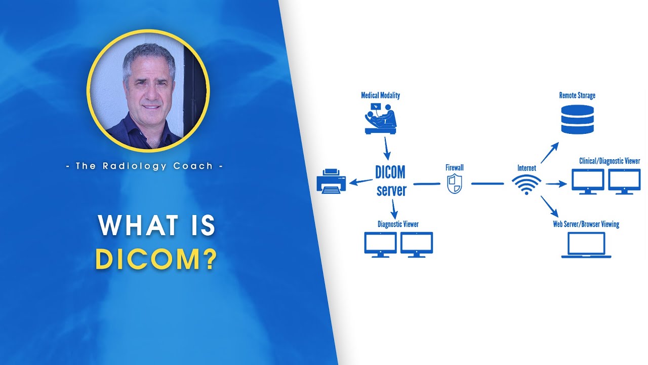
-DICOM stands for Digital Imaging and Communications in Medicine. It is a standard protocol for the transmission, sharing, and archiving of medical images and related information, such as patient data and study details. It allows for the digital processing and transfer of images from the point of care to the radiologist's display monitor.
Who were the key figures in the discovery and development of X-rays and radiation?
-William Rankin discovered the use of X-rays in 1895. Physicists Marie Curie, Pierre Curie, and Henri Becquerel were awarded the Nobel Prize in 1903 for their discovery of radiation. Their work has significantly influenced the field of medical imaging.
How were X-rays processed and viewed in the past before digitalization?
-In the past, X-rays were printed onto films that needed to be processed in a dark room, washed, dried, and then hung against a light source for radiologists to view. This process was time-consuming and required physical handling of the films.
What is the difference between a PACS and a VNA in the context of medical imaging?
-A PACS (Picture Archiving and Communication System) is a digital solution for the storage, transfer, and viewing of medical images, primarily used in radiology departments. A VNA (Vendor Neutral Archive), on the other hand, is a more comprehensive system designed to store, transmit, and view all types of medical images from various departments, regardless of the vendor or image format.
What is a 'hanging protocol' in the context of X-ray films?
-A hanging protocol refers to the deliberate arrangement of X-ray films against a light source based on the type of exam performed. This arrangement helps radiologists efficiently review and interpret the images during a study.
How are annotations made on digital X-ray images in a PACS system?
-In a PACS system, annotations such as measurements and arrows are made digitally on the images. These annotations are stored as part of the PACS system and can be viewed by other users, enhancing the collaborative aspect of medical image analysis.
What is the significance of DICOM tags in organizing patient files?
-DICOM tags are metadata elements within a DICOM file that contain specific information such as patient name, date of service, and other demographic details. They are crucial for organizing and searching patient files within a PACS or VNA system.
How does a VNA handle non-DICOM images?
-A VNA handles non-DICOM images by utilizing an XDS (Cross-Enterprise Document Sharing) registry to store metadata instead of DICOM tags. This allows the VNA to manage and retrieve images that do not conform to the DICOM standard.
What is the role of NEMA in the DICOM standard?
-NEMA (National Electrical Manufacturers Association) is responsible for determining the specific categories or 'classes' that medical images must fall into within the DICOM standard. This ensures consistency and interoperability across different medical imaging devices and systems.
Why is the transition to digital imaging important in medical settings?
-The transition to digital imaging is important because it allows for faster image transfer, easier storage and retrieval, and more efficient collaboration among healthcare professionals. It also reduces the risk of physical damage to images and enhances the overall quality of patient care.
Outlines

Dieser Bereich ist nur für Premium-Benutzer verfügbar. Bitte führen Sie ein Upgrade durch, um auf diesen Abschnitt zuzugreifen.
Upgrade durchführenMindmap

Dieser Bereich ist nur für Premium-Benutzer verfügbar. Bitte führen Sie ein Upgrade durch, um auf diesen Abschnitt zuzugreifen.
Upgrade durchführenKeywords

Dieser Bereich ist nur für Premium-Benutzer verfügbar. Bitte führen Sie ein Upgrade durch, um auf diesen Abschnitt zuzugreifen.
Upgrade durchführenHighlights

Dieser Bereich ist nur für Premium-Benutzer verfügbar. Bitte führen Sie ein Upgrade durch, um auf diesen Abschnitt zuzugreifen.
Upgrade durchführenTranscripts

Dieser Bereich ist nur für Premium-Benutzer verfügbar. Bitte führen Sie ein Upgrade durch, um auf diesen Abschnitt zuzugreifen.
Upgrade durchführenWeitere ähnliche Videos ansehen


5.0 / 5 (0 votes)
