T2 Relaxation, Spin-spin Relaxation, Free Induction Decay, Transverse Decay | MRI Physics Course #4
Summary
TLDRThis educational video script delves into the intricacies of nuclear magnetic resonance (NMR) and relaxation processes in MRI. It explains how protons align in an external magnetic field and resonate at a specific frequency. The script then focuses on T2 relaxation, detailing the loss of transverse magnetization due to spin-spin interactions and the impact of magnetic field inhomogeneities. It uses the analogy of spinning basketballs to illustrate dephasing and discusses T2 star decay, highlighting the differences between T2 and T2 star relaxation times. The script also explores how T2 relaxation curves vary across tissues and the role of echo time (TE) in MRI imaging, emphasizing its importance in generating tissue contrast.
Takeaways
- 🧲 The process of nuclear magnetic resonance (NMR) involves placing protons in an external magnetic field, causing them to align and resonate at a specific frequency.
- 🌀 A radiofrequency (RF) pulse is applied perpendicular to the magnetic field, causing protons to resonate in phase and fan out from the longitudinal magnetization vector.
- 🔄 At 90 degrees, the net magnetization vector transitions from longitudinal to transverse, resulting in maximum transverse magnetization.
- 📉 T2 relaxation refers to the loss of transverse magnetization, which occurs due to dephasing of spins as the RF pulse is stopped.
- 🏀 Spin-spin relaxation, characterized by the dephasing of spins due to interactions, is a key component of T2 relaxation, with the rate depending on the tissue type.
- 🌐 T2 star decay accounts for the loss of signal due to both spin-spin interactions and magnetic field inhomogeneities, which are not solely due to spin interactions.
- 🛑 The MRI scanner's inability to create a perfectly homogeneous magnetic field contributes to T2 star decay, along with the presence of substances in the patient and local magnetic field disruptions.
- 🔄 A 180-degree RF pulse can be used to rephase spins that have dephased, effectively compensating for magnetic field inhomogeneities and recovering the T2 relaxation signal.
- 📊 T2 relaxation curves vary by tissue type, with water or CSF showing less dephasing and thus slower signal loss compared to fat, which has more rapid signal decay.
- ⏱ Adjusting the time to echo (TE) in MRI can manipulate the contrast between tissues by highlighting differences in T2 relaxation times, allowing for clearer differentiation in imaging.
Q & A
What is nuclear magnetic resonance (NMR)?
-Nuclear magnetic resonance is a process where protons within an external magnetic field align with that field and resonate at a set frequency. A radiofrequency magnetic pulse is applied, causing the protons to start resonating in phase with one another, leading to a loss of longitudinal magnetization and a gain of transverse magnetization.
What are the two types of relaxation processes discussed in the script?
-The two types of relaxation processes discussed are T2 relaxation, which is the loss of transverse magnetization, and T1 relaxation, which is the regaining of longitudinal magnetization.
What is meant by 'spin-spin relaxation'?
-Spin-spin relaxation refers to the loss of transverse magnetization that occurs as protons or spins go out of phase with one another after the radio frequency pulse is stopped. This dephasing is primarily caused by interactions between the spins themselves.
How does the composition of fat affect its T2 relaxation?
-Fat, composed of long chains of triglycerides, allows the molecules to bump into each other easily, leading to more frequent interactions between spins. This results in a quicker loss of phase and faster T2 relaxation, causing a rapid decrease in signal compared to tissues like CSF.
What is the significance of the T2 star decay in MRI?
-T2 star decay in MRI accounts for the loss of signal due to both spin-spin interactions and magnetic field inhomogeneities. It is a measurable decay that occurs more quickly in tissues than the ideal T2 decay, which is solely due to spin-spin interactions.
Why do magnetic field inhomogeneities affect the T2 star decay?
-Magnetic field inhomogeneities affect the T2 star decay because they cause protons at different locations to experience varying magnetic field strengths, leading to different precession rates and increased dephasing, which in turn accelerates the loss of transverse magnetization.
How does the 180-degree radio frequency pulse help in compensating for magnetic field inhomogeneities?
-The 180-degree radio frequency pulse refocuses the dephasing spins by flipping their phase, allowing the faster and slower precessing spins to realign. This effectively compensates for the local field inhomogeneities, leading to a re-phasing of the spins and an increase in the transverse magnetization vector.
What is the purpose of the time to echo (TE) in MRI?
-The time to echo (TE) in MRI is the time interval between the 90-degree RF pulse and the point at which the signal is sampled. It is crucial for controlling the contrast in the image by highlighting differences in T2 relaxation times between various tissues.
How does the choice of TE affect the MRI image contrast?
-A shorter TE results in a higher signal but less contrast between tissues, as the differences in T2 relaxation times are not fully expressed. A longer TE allows for greater distinction between tissues based on their T2 relaxation properties, increasing the contrast in the image.
What is the relationship between T2 relaxation and the appearance of different tissues in MRI?
-T2 relaxation times vary between different tissues, with some tissues like CSF having longer T2 times and appearing brighter, while others like muscle have shorter T2 times and appear darker. The MRI image contrast is directly influenced by these T2 differences.
Outlines
🧲 Understanding Nuclear Magnetic Resonance and T2 Relaxation
The paragraph introduces the concept of nuclear magnetic resonance (NMR), where protons align with an external magnetic field and resonate at a specific frequency. A radiofrequency pulse is applied to cause the protons to resonate in phase, leading to a loss of longitudinal magnetization and a gain in transverse magnetization. The lecture focuses on T2 relaxation, which is the loss of transverse magnetization. Spin-spin relaxation is explained as the process where protons lose phase coherence due to interactions with each other, leading to a decrease in transverse magnetization. The analogy of spinning basketballs is used to illustrate how spins can go out of phase when they interact with one another. The paragraph also discusses the difference between T2 relaxation and T2* decay, with the latter including signal loss due to magnetic field inhomogeneities. Examples of fat and CSF tissues are used to demonstrate how the rate of signal loss varies between different types of tissues due to their molecular structures and interactions.
🌀 Exploring T2* Decay and Its Causes
This paragraph delves deeper into T2* decay, which is the measurable decay of the transverse magnetization vector in MRI. It is distinguished from T2 decay by the fact that T2* includes signal loss due to magnetic field inhomogeneities, not just spin-spin interactions. The paragraph explains that T2* decay occurs more quickly in tissues due to these inhomogeneities. Three mechanisms causing magnetic field inhomogeneities are discussed: imperfections in the MRI scanner's magnetic field, substances within the patient that disrupt local magnetic fields, and the de-phasing of spins themselves. The paragraph emphasizes the importance of understanding T2* decay for imaging, as it affects the contrast and visibility of different tissues in MRI scans.
🔄 Compensating for T2* Decay with 180-Degree RF Pulse
The paragraph explains how the T2* decay, caused by local magnetic field inhomogeneities, can be compensated for in MRI imaging. It describes the process of applying a 180-degree radiofrequency (RF) pulse to re-phase the de-phasing spins, allowing for a more accurate measurement of T2 relaxation. The use of a 180-degree RF pulse effectively 'refocuses' the spins, leading to a temporary increase in the transverse magnetization vector. This technique is crucial for generating an echo, which is a signal that can be sampled to provide information about T2 relaxation. The paragraph also touches on the concept of time to echo (TE), explaining how varying this time can affect the signal strength and the contrast between different tissues in an MRI image.
📊 Impact of Time to Echo on MRI Contrast
This final paragraph discusses the practical implications of the time to echo (TE) on MRI imaging. It explains how different TE values can be chosen to highlight the T2 relaxation differences between tissues, which in turn affects the contrast in the resulting MRI images. The paragraph illustrates how a short TE results in high signal but low contrast between tissues, while a longer TE increases contrast by showing more significant differences in signal intensity. The importance of selecting the appropriate TE for imaging specific tissues is emphasized, as it directly influences the visibility and diagnostic utility of the MRI scan. The paragraph concludes by setting the stage for the next lecture, which will cover T1 relaxation and its implications for MRI imaging.
Mindmap
Keywords
💡Nuclear Magnetic Resonance (NMR)
💡Magnetization Vector
💡Transverse Magnetization
💡T2 Relaxation
💡Spin-Spin Relaxation
💡T2 Star Decay
💡Magnetic Field Inhomogeneities
💡Echo
💡Time to Echo (TE)
💡Contrast
Highlights
Nuclear magnetic resonance (NMR) involves placing protons in an external magnetic field, causing them to align and resonate at a set frequency.
A radiofrequency magnetic pulse is applied perpendicularly to initiate proton resonance and fanning out from the longitudinal magnetization vector.
At 90 degrees, the net magnetization vector reaches maximum transverse magnetization, losing longitudinal magnetization.
Relaxation processes are discussed, including T2 relaxation (loss of transverse magnetization) and T1 relaxation (regaining longitudinal magnetization).
T2 relaxation is the focus of the lecture, detailing the loss of transverse magnetization due to spin-spin interactions.
Spin-spin relaxation is characterized by protons going out of phase with each other, influenced by the tissue they're in.
Transverse Decay is introduced as the loss of net transverse magnetization vector due to proton dephasing.
An analogy of spinning basketballs is used to explain the dephasing process in T2 relaxation.
Fat and CSF are compared to illustrate how their molecular structures affect the rate of transverse magnetization loss.
T2 relaxation curves are plotted to show how different tissues lose phase at varying rates.
T2 star Decay is distinguished from T2 Decay, accounting for magnetic field inhomogeneities.
The concept of T2 star time constants is introduced, explaining the time it takes for 63% signal loss.
Three mechanisms causing magnetic field inhomogeneities are discussed: MRI scanner limitations, patient substances, and local spin dephasing.
T2 star Decay is shown to be influenced by the magnetic field strength experienced by protons at different locations.
A 180-degree radio frequency pulse is introduced as a method to rephase protons and compensate for local magnetic field inhomogeneities.
The echo mechanism in MRI is explained, showing how it can be used to regain T2 relaxation and account for magnetic field inhomogeneities.
The impact of time to echo (TE) on MRI signal and tissue contrast is discussed, showing how it can be manipulated to highlight T2 relaxation differences.
The lecture concludes with a preview of the next topic, T1 relaxation, and its implications for imaging tissue differences.
Transcripts
hello everybody and welcome back so in
the previous talk we looked at the
process of nuclear magnetic resonance
where we placed protons within an
external magnetic field and they aligned
with that magnetic field processing at a
set frequency we then applied a
perpendicular radiofrequency magnetic
pulse that caused those protons to start
resonating in Phase with one another and
Fanning out away from that longitudinal
magnetization Vector that net
magnetization Vector then gained more
and more transverse magnetization and if
we flip that net magnetization Vector to
90 degrees we completely lost the z-axis
magnetization the longitudinal
magnetization and at the same point
we've completely gained transverse
magnetization at 90 degrees we have our
maximum transverse magnetization Vector
so that process of nuclear magnetic
resonance and the application of a radio
frequency pulse caused loss of
longitudinal magnetization and gain of
transverse magnetization in the next two
talks we're going to look at the process
of relaxation which happens in two
separate independent mechanisms the
first is the loss of transverse
magnetization otherwise known as T2
relaxation the second process which is
independent of that first process is
what's known as T1 relaxation or the
gaining regaining of longitudinal
magnetization so in today's lecture
we're going to look at T2 relaxation
which is the loss of transverse
magnetization now there are multiple
different terms for this that I want to
introduce you to the first is what's
known as spin spin relaxation we've seen
in previous talks that the loss of
transverse magnetization comes from this
spins going out of phase with one
another as we stop that radio frequency
pulse the spins that were resonating in
Phase start to de-phase from one another
and the rate at which they defaze
depends on which tissue they're in now
that dephasing is primarily caused by
spins interacting with one another the
way I to remember this is that spin spin
has two s's here so spin spin is related
to T2 relaxation
now if you take that analogy of spinning
a basketball on your finger if I had
many people in the room and everyone was
spinning a basketball on their finger
and the basketballs bumped into one
another those basketballs will start to
lose some of their Spin and they will be
de-phasing or spinning at different
rates from the basketballs in the room
that is the process for the loss of
phase in T2 relaxation another term that
you may come across is what's known as
transverse Decay and this makes sense as
protons defaze they lose their
transverse or their net transverse
magnetization vector and we get a loss
of signal because it's a transverse
signal that we're measuring in our MRI
machine
so if you imagine these as basketballs
these protons here and they're bouncing
into one another and as the spins
interact with one another the spins with
different energy levels as well the
energy is transferred and those spins
become out of phase and that's the
predominant mechanism for the loss of
transverse magnetization so let's have a
look at an example here we have a unit
of fat represented by this orange color
here and a unit of CSF represented by
this blue color here we've applied a
radio frequency pulse that matches the
processional frequency of these net
magnetization vectors and we flip that
net magnetization Vector to 90 degrees
our maximum signal now what happens when
we turn off that radio frequency pulse
while those processing spins will now
start to de-phase and if you think about
what fat is made up of it's made up of
long chains of triglycerides where all
the molecules are joined together they
can bump into each other really easily
if we take our room full of people with
basketballs on their hands fat has got
chains of people holding hands with one
another and as they move around the room
they're much more likely to have their
spins or the basketballs bump into one
another
water or CSF has free people walking
around in the room free to move as they
please they're not joined to other
people in these long chains of fatty
acids so water they're less likely for
the spins to interact with one another
there's more free movement in the room
so you'll see that the phase of water or
CSF stays much more in Phase than fat
does let's have a look at these two
separate tissues and see how they behave
differently as they start to lose phase
in the transverse plane
as you see in CSF here the net
magnetization Vector is staying much
more in Phase the spins aren't
interacting as much as they are in
fattier and we see that the signal
generated from fat is lost much more
quickly than the signal generated from
CSF
so now we can draw these curves here
which are our T2 relaxation curves that
are dependent on the type of tissue
through which the spins are spinning
look in fact here how outer phase those
spins are and as we know as we get out
of phase our magnetization vectors in
the X Y plane start to cancel each other
out and after a period of time we're
getting complete loss of that signal in
fat the water has stayed relatively in
Phase with one another and although
we're still getting lots of signal
because they're not perfectly in Phase
that loss is much slower and for each
tissue we can plot that free induction
Decay curve for the different tissues
now you'll see that I've written T2 star
Decay here and not T2 Decay now whenever
you put an asterisk somewhere it means
that there's some terms and conditions
and the reason for this T2 star Decay is
because the actual measurable Decay that
we measure on the MRI machine the drop
off of signal that we were looking at at
the previous slide is not purely due to
the spins interacting with one another
T2 relaxation in an ideal world would
only be getting loss of that transverse
magnetization Vector from spins
interacting with one another spin spin
relaxation now in the real world we get
loss of signal because of spin spin
relaxation but we also get loss of
signal due to magnetic field in
homogeneities which we're going to look
at next before we move into that I want
to draw your attention to these T2 star
time constants here
at the beginning of our radio frequency
pulse once we flip those net
magnetization vectors to 90 degrees we
have maximum signal and all those
protons are in Phase with one another we
have a hundred percent of the transverse
signal at 90 degrees this is our
transverse magnetization vector
now as time goes by we get lots of that
signal because of the dephasing of those
atoms and it happens at different rates
depending on the tissue we're in
when 63 of that signal has been lost or
we have 37 of the transverse
magnetization Vector left that time
constant the amount of time it takes to
get to that point is what's known as T2
star or T2 star Decay and we can use
these values to get contrast in our
tissues later
now in an Ideal World we wouldn't want
T2 star we would want a T2 value which
would represent 63 loss in the
transverse magnetization Vector purely
due to spin spin interactions not due to
magnetic field in homogeneities and this
curve here would be known as our T2
relaxation curve we've seen that the t2
star relaxation curve happens much
quicker in tissues now if we have a look
at our MRI machine here in an Ideal
World the magnetic field would be
homogeneous it would be exactly the same
no matter where the protons are within
this magnetic field now there are three
separate mechanisms that make this
magnetic field inhomogeneous and cause
that T2 star Decay the first is that the
MRI scanner itself can't make a perfect
strength magnetic field that's equal all
the way through the transverse plane the
coils are going to have differing
magnetic field strains the further away
from the coils you get so that's the
first reason for magnetic field in a
motor geneties the second mechanism is
that there could be a substance within
the patient either metal or calcium or
dense cortical bone that causes
disruption in the local magnetic fields
here and that's why in a patient that
has a metal device you'll often see T2
signal is completely lost around that
device that's because of the localized
changes in the magnetic field strength
and the last thing is when spins start
to de-phase with one another the
magnetization vectors are becoming out
of phase with one another and they can
disrupt the local magnetic field as well
and so we don't get perfect magnetic
field lines in the longitudinal plane
here so all three of those mechanisms
cause the magnetic field to be in
homogeneous now because the field is
inhomogeneous a proton that is sitting
here will experience a different
magnetic field strength to a proton that
is sitting at a different location and
we've seen that when protons or spins
experience different magnetic field
strands they will spin at different
rates we look back to our alarm
frequency a different magnetic field
strength will cause the dephasing to be
increased because the rates of change of
those processional values will be
different between those two protons and
that's what's responsible for this T2
star effect occurring now our T2 star or
the free induction Decay Curve will
always be less than the t2 value the t2
relaxation value and in imaging we want
to try and compensate for this reduction
or increased rate of loss of transverse
magnetization and there actually is a
mechanism for which we can compensate
for these local field in homogeneous 80s
so let's have a look at how we go about
compensating for that T2 star decay when
we are trying to produce an image the
first thing we need to do is apply a 90
degree RF pulse that is perpendicular to
the main magnetic field once we've
applied that 90 degree RF pass and turn
it off we will get relaxation T2
relaxation where we get loss of
transverse magnetization now in an Ideal
World the transverse magnetization loss
will be June only due to spin spin
interactions where spins are
transferring energy and they start
becoming out of phase because of that
transfer of energy between the two spins
that is what's known as our T2 Decay or
T2 relaxation
now what actually happens in the real
world is we get spin spin interactions
which cause that loss of transverse
magnetization and we get local magnetic
field in homogeneities which causes this
T2 star Decay to occur so this is what
we want this T2 relaxation this is what
we actually measuring because of the
inhomogeneities within the magnetic
field
so what has actually happened here well
we've taken our longitudinal net
magnetization vector and flipped it to
90 degrees with that 90 degree RF pulse
we've completely lost longitudinal
magnetization and we've now got a
maximum transverse magnetization we've
got maximum T2 signal here now what's
going to happen is these spins in the
same voxel within our image are going to
defaze with one another if we look at it
end on they're going to D phase like
this some of them will move faster than
others and that's mainly due to the spin
spin interaction between the different
spins but we've also seen that there is
differing strengths of magnetic field
strength because of that local
inhomogeneity in the magnetic field now
the one that's experiencing a higher
magnetic field strength is going to
defaze quicker than the one that's
experiencing a lower magnetic field
strength so we've flipped it to 90
degrees and we're getting de-phasing of
these spins
now what happens is over time these
spins will defaze with one another and
they will also start gaining
longitudinal magnetization now the one
is dephasing faster than the other what
we want to do is be able to re-phase
these two spins with one another and the
way we do that is by applying what's
known as a 180 degree radio frequency
pulse it's the same radio frequency
pulses this 90 degree radio frequency
pulse same magnitude but for twice the
duration so what has happened now one
spin is de-phasing faster than another
Spin and we apply a 180 degree RF pole
so let me get this right here this is
the faster one the blue is the slower
one we apply a 180 degree radio
frequency pulse we are flipping those
spins now 180 degrees here is our main
magnetic field
now the leading spin is the slow Spin
and the trailing spin is the fast spin
we're spinning or processing in this
direction now what is going to happen
over time is the faster spin is going to
catch up with the slower or the lagging
Spin and if we wait the exact same
period of time between our 90 and 180
degree pulse what will happen is those
spins will now become in Phase with one
another because of that 180 degree spoon
and we have gained now that net
magnetization Vector in the transverse
plane our spins have re-phased with one
another and you can see that represented
by this graph here is that as the spins
start to re-phase with one another we
get an increase in that transverse
magnetization Vector because of that 180
degree flip and then allowing those
spins now to catch up with one another
and sync up giving us a maximum net
transverse magnetization vector and what
we can do then is sample the signal at
this point and if we sample the signal
at this point you'll notice that that
signal is the same as the t2 relaxation
the signal we're measuring now at the
time to Echo and you can see now why
it's called an echo is the same as what
we would have gotten if the loss of
transverse magnetization was only
because of spin to spin interactions or
T2 relaxation and it's this mechanisms
which we're going to look at later in a
pulse sequence called spin Echo
sequences that allows us to regain that
T2 relaxation and account for those
local inhomogeneities in the magnetic
field and we can do this for all the
different tissues all the different
voxels within our patient and plot these
values over time now importantly we can
place this 180 degree RF pulse wherever
we want to place it and then measure the
echo at the same distance between the 90
degree RF pulse and the 180 degree RF
boss this distance and this distance is
the same we can make this te time much
shorter or or we can make it much longer
a shorter te time will give us higher
signal and a longer te time is going to
give us lower signal and if we plot
those signals over time depending on the
different tissues that we're trying to
image we can see that T2 relaxation
curve it takes much longer in CSF
because those hydrogen protons are able
to move freely in fact you think of
people holding hands in the room
spinning basketballs in those long
triglyceride change the basketballs are
going to bump into each other and that
spin spin interaction is going to cause
a loss of transverse magnetization and
that happens even faster in muscle
now we've seen that we can choose the
time to Echo when we're going to sample
this tissue if we sample really early a
short time to Echo we flip that
longitudinal magnetization Vector into
90 degrees switch off the RF pulse and
immediately sample the tissue what we
get is a short time to Echo now you'll
see that the signal here is high for the
muscle for the fat and for the CSF we're
going to have a high signal and there's
going to be no contrast between these
tissues we've got very little difference
in the t2 relaxation times between these
tissues if we wait a longer period of
time and make our Echo slightly longer
you'll see that the signal has decreased
but the contrast between the various
different tissues has increased our
muscle signal is going to be much lower
it's going to be represented darker on
the MRI fat is lower than CSF but higher
than muscle and now CSF is still giving
us a bright signal value waiting or
prolonging the t2 times going to
increase the contrast between those two
tissues so you can see how changing te
time changes the contrast and that
contrast is based on the t2 relaxation
differences between these tissues now we
can wait even longer and have a third
time to Echo here where we've now got
very little signal and again we've lost
contrast here there's very slight
grayscale differences but now it's
difficult to tell the CSF from the fat
and from the muscle so if you wait too
long without time to Echo we're going to
lose that transverse magnetization
vector and not have any signal to detect
now hopefully this graph shows you that
change in the te will highlight the
differences in T2 relaxation differences
between the various different types of
tissues in the next talk we're going to
be looking at T1 relaxation and I'm
going to show you how we can use T1
relaxation differences in order to see
the T1 differences between tissues so
until that talk I'll see you all then
goodbye everybody
تصفح المزيد من مقاطع الفيديو ذات الصلة
T1 Relaxation, Spin-lattice Relaxation, Longitudinal Recovery | MRI Physics Course #5
MRI Physics | Magnetic Resonance and Spin Echo Sequences - Johns Hopkins Radiology
T1, T2 and Proton Density Weighting | MRI Weighting and Contrast | MRI Physics Course #6
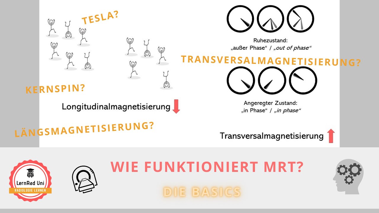
Wie funktioniert MRT? Die Basics
Wie funktioniert MRT? T1 und T2 Wichtung
Spin, Precession, Resonance and Flip Angle | MRI Physics Course | Radiology Physics Course #3
5.0 / 5 (0 votes)