BIOLOGI SMA Kelas 11 - Sistem Pernapasan | GIA Academy
Summary
TLDRThis educational video from Gia Academy explores the human respiratory system, detailing the organs involved in breathing and their functions. It explains the process of inhaling oxygen and exhaling carbon dioxide and water vapor, highlighting the roles of the nasal cavity, pharynx, larynx, trachea, bronchi, bronchioles, and alveoli. The video also covers the mechanics of breathing, including diaphragm and intercostal muscle actions, and discusses various respiratory diseases. It concludes with a look at the different volumes of air involved in breathing and factors affecting respiration rates.
Takeaways
- 😷 The human respiratory system includes organs such as the nasal cavity, pharynx, larynx, trachea, bronchi, bronchioles, alveoli, and lungs.
- 👃 The nasal cavity filters, warms, and humidifies the air, and also detects scents through the olfactory receptors.
- 🔄 The pharynx serves as a junction between the nasal cavity and the respiratory and digestive tracts, with the epiglottis preventing food from entering the windpipe.
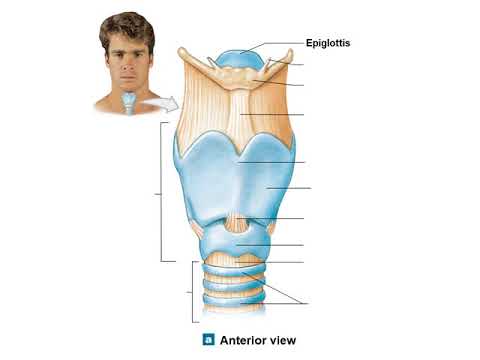
- 🗣️ The larynx contains the vocal cords and the epiglottis, which plays a crucial role in both breathing and swallowing.
- 🌀 The trachea, or windpipe, is a tube that branches into the lungs and is lined with cartilage rings to keep it open.
- 💨 Bronchi and bronchioles are the branching airways within the lungs that end in tiny air sacs called alveoli.
- 🌪️ Alveoli are the site of gas exchange, where oxygen diffuses into the bloodstream, and carbon dioxide diffuses out.
- 🏋️♂️ Breathing is facilitated by respiratory muscles, including the intercostal muscles and the diaphragm, which expand and contract the chest cavity.
- 🔁 The process of inhalation (inspiration) and exhalation (expiration) involves the exchange of oxygen and carbon dioxide, regulated by changes in air pressure within the thoracic cavity.
- 🚑 Common respiratory disorders include asthma, emphysema, lung cancer, pneumonia, and tuberculosis, which can impair the function of the respiratory system.
Q & A
What is the primary function of the respiratory system?
-The primary function of the respiratory system is to facilitate the intake of oxygen into the body and the expulsion of carbon dioxide and water vapor from the body.
What are the main organs that make up the human respiratory system?
-The main organs that make up the human respiratory system include the nasal cavity, pharynx, larynx, trachea, bronchi, bronchioles, alveoli, and lungs.
How does the nasal cavity contribute to the respiratory process?
-The nasal cavity contributes to the respiratory process by filtering, warming, and humidifying the air that enters the body, and it also detects odors through the olfactory receptors.
What is the role of the epiglottis in the respiratory system?
-The epiglottis plays a crucial role in preventing food from entering the trachea during swallowing by covering the entrance to the larynx.
How does the trachea contribute to the respiratory process?
-The trachea, or windpipe, serves as a conduit for air to travel from the upper airway to the bronchi, which then branch out into the lungs.
What is the function of the bronchi and bronchioles in the respiratory system?
-The bronchi and bronchioles are the branching airways in the lungs that eventually lead to the alveoli, where gas exchange occurs.
What is the significance of the alveoli in the respiratory system?
-Alveoli are the tiny air sacs in the lungs where the actual gas exchange between oxygen and carbon dioxide takes place, allowing oxygen to enter the bloodstream and carbon dioxide to be expelled.
How does the diaphragm muscle assist in the breathing process?
-The diaphragm muscle assists in the breathing process by contracting and relaxing, which changes the volume of the thoracic cavity, thus facilitating inhalation and exhalation.
What is the difference between external and internal respiration?
-External respiration refers to the gas exchange that occurs in the alveoli of the lungs, while internal respiration refers to the gas exchange that occurs within the body's tissues and cells.
What is the role of hemoglobin in the respiratory system?
-Hemoglobin plays a critical role in the respiratory system by binding to oxygen to form oxyhemoglobin, which transports oxygen from the lungs to the body's tissues.
What are some common respiratory disorders mentioned in the script?
-Some common respiratory disorders mentioned in the script include asphyxiation, asthma, emphysema, lung cancer, pneumonia, tuberculosis, influenza, and bronchitis.
Outlines
🌬️ Introduction to the Respiratory System
The script begins with a welcome and an introduction to the topic of respiration. It explains that breathing is the process of taking in oxygen and expelling carbon dioxide and water vapor. The human respiratory system is composed of various organs including the nasal cavity, pharynx, larynx, trachea, bronchi, bronchioles, and alveoli. Each of these organs plays a specific role in the respiratory process. For instance, the nasal cavity filters, moistens, and warms the air, while the larynx houses the vocal cords and epiglottis which prevent food from entering the windpipe. The trachea, shaped like a pipe, branches into the bronchi within the lungs, and the bronchi further divide into bronchioles. The alveoli are the site of gas exchange where oxygen diffuses into the blood, and carbon dioxide diffuses out.
🔁 Breathing Mechanics and Gas Exchange
This section delves into the mechanics of breathing, detailing the process of inspiration (inhalation) and expiration (exhalation). It explains how changes in air pressure within the thoracic cavity, diaphragm, and abdominal cavity regulate these processes. The respiratory muscles, including the intercostal muscles and the diaphragm, are highlighted for their roles in expanding and contracting the thoracic cavity to facilitate breathing. The script also discusses two types of respiration: chest (diaphragmatic) and abdominal, each involving different muscles and mechanisms. Furthermore, it describes the external and internal respiration processes, where external respiration occurs in the alveoli with oxygen binding to hemoglobin, and internal respiration involves oxygen diffusing into body tissues and cells for cellular respiration, producing energy, carbon dioxide, and water.
🌡️ Regulation of Breathing and Respiratory Disorders
The script discusses how the body regulates the production and expulsion of carbon dioxide, which is a critical aspect of maintaining acid-base balance in the blood. It outlines the various forms in which carbon dioxide is transported in the blood, including as dissolved carbon dioxide, carbaminohemoglobin, and bicarbonate ions. The section also covers the different volumes of air involved in breathing, such as tidal volume, inspiratory reserve volume, expiratory reserve volume, residual volume, vital capacity, and total lung capacity. Additionally, factors influencing respiratory rate, such as age, gender, body position, temperature, and activity level, are mentioned. The paragraph concludes with a discussion on various respiratory disorders, including asphyxiation, asthma, emphysema, lung cancer, pneumonia, tuberculosis, influenza, and others, which can impair the respiratory system and affect overall health.
📚 Summary and Quiz on Respiratory System
The final part of the script provides a summary of the respiratory system's structure, function, and the mechanics of breathing. It also presents a quiz to test the understanding of the audience regarding internal respiration processes, the mechanics of chest breathing, and the identification of different volumes of air during respiration. The quiz includes questions on identifying the correct processes associated with internal respiration, determining the mechanism of breathing from a given illustration, and identifying the type of air volume described in a scenario. The script ends with a reminder to continue learning about the respiratory system through further videos and to look forward to the next video in the series.
Mindmap
Keywords
💡Respiration
💡Respiratory System
💡Alveoli
💡Diaphragm
💡Intercostal Muscles
💡Inspiratory Reserve Volume
💡Expiratory Reserve Volume
💡Residual Volume
💡Vital Capacity
💡Respiratory Rate
💡Pneumonia
Highlights
The video introduces the human respiratory system, explaining how oxygen enters and carbon dioxide exits the body.
The respiratory system consists of several organs: the nasal cavity, pharynx, larynx, trachea, bronchi, bronchioles, alveoli, and lungs.
The nasal cavity plays a key role in filtering air, regulating temperature, and detecting smells through olfactory nerves.
The pharynx serves as a crossroad between the respiratory and digestive systems, allowing air to move to the trachea and food to the esophagus.
The larynx, also known as the voice box, contains vocal cords and the epiglottis, which prevents food from entering the windpipe during swallowing.
The trachea, or windpipe, is a tube made up of cartilage rings that transport air to the bronchi. It has three layers: outer connective tissue, middle smooth muscle, and inner ciliated epithelium.
Bronchi are two branches that extend from the trachea to the lungs, which further divide into bronchioles, smaller airways without cartilage.
Alveoli, small sac-like structures, are responsible for gas exchange between oxygen and carbon dioxide. Their thin walls facilitate diffusion, supported by capillaries.
The lungs are the main respiratory organs, divided into lobes: three in the right lung and two in the left. They are surrounded by a pleura membrane and fluid to reduce friction.
Breathing involves inspiration (inhaling oxygen) and expiration (exhaling carbon dioxide). This occurs 15-18 times per minute under normal conditions.
Breathing can be classified into two types: chest breathing (intercostal muscles) and abdominal breathing (diaphragm muscles).
External respiration occurs in the alveoli, where oxygen enters the blood and carbon dioxide exits, while internal respiration happens in the tissues, exchanging gases between blood and cells.
Hemoglobin plays a key role in transporting oxygen by forming oxyhemoglobin. Oxygen is released in tissues during internal respiration for cellular respiration.
Diseases of the respiratory system include asthma, emphysema, lung cancer, pneumonia, tuberculosis, and bronchitis, which impair lung function.
Factors such as age, gender, activity level, and temperature can influence breathing rate, with faster rates seen in younger individuals, females, and during increased physical activity.
Transcripts
[Musik]
Halo teman-teman selamat datang kembali
di Gia Academy youtube channel Semoga
teman-teman selalu sehat dan terus
semangat
setiap waktu kita bernafas menghirup
udara segar kita masukkan udara lewat
hidung kemudian menghembuskannya kembali
lewat hidung atau mulut kira-kira
Seperti apa ya proses saat udara masuk
dan keluar dari tubuh kita kita akan
mempelajarinya di video kali ini
jadi di video ini kita akan belajar
tentang sistem pernapasan simak terus
videonya ya
nah teman-teman seperti yang kita tahu
bernafas adalah proses memasukkan
oksigen ke dalam tubuh dan mengeluarkan
karbondioksida dan uap air dari tubuh
sistem pernapasan manusia terdiri dari
organ-organ pernapasan yaitu rongga
hidung faring laring tenggorokan atau
trakea bronkus bronkiolus alveolus dan
paru-paru Mari kita bahas struktur dan
fungsinya satu persatu
[Musik]
pertama rongga hidung rongga hidung
merupakan tempat masuknya udara dari
luar rongga hidung selalu lembab karena
adanya lendir di dalam rongga hidung
juga terdapat tulang hidung Serabut
saraf pembau atau olfaktori Serabut
saraf menuju otak dan rambut-rambut
halus atau silia saraf pembau selaput
lendir dan silia berfungsi menyaring
debu dan kotoran yang masuk ke hidung
mengatur suhu udara dan mengenali bau
melalui saraf olfaktori
[Musik]
organ pernapasan kedua faring atau teka
udara dari rongga hidung akan masuk ke
faring faring merupakan persimpangan
antara rongga hidung ke tenggorokan atau
saluran pernapasan dan rongga hidung ke
kerongkongan atau saluran pencernaan
Pada bagian belakang faring terdapat
laring
berikutnya laring pada laring terdapat
pita suara dan epiglotis atau katup
pangkal tenggorokan pada waktu menelan
makanan epiglotis menutupi laring
sehingga makanan tidak masuk ke
tenggorokan sementara itu pada waktu
bernapas epiglotis akan membuka sehingga
udara masuk ke laring lalu menuju
tenggorokan di bagian bawah epiglotis
terdapat tulang rawan tiroid yang
membentuk jakun yang lebih tampak pada
laki-laki daripada wanita
selanjutnya trakea atau batang
tenggorokan trakea berbentuk seperti
pipa dengan panjang kurang lebih 10 cm
trakea bercabang dua dalam paru-paru
membentuk bronkus dinding trakea Terbagi
3 yaitu lapisan terluar terdiri dari
jaringan ikat lapisan Tengah terdiri
dari otot polos dan cincin tulang rawan
trakea tersusun dari
16-20 cincin tulang rawan yang berbentuk
huruf C bagian belakang cincin tulang
rawan tidak tersambung ke esofagus
sehingga menyebabkan trakea tetap
terbuka Lapisan dalam terdiri dari
jaringan epitel silindris selapis
bersilia yang menghasilkan lendir lendir
berfungsi menangkap debu dan
mikroorganisme yang masuk saat menghirup
udara selanjutnya debu dan
mikroorganisme didorong oleh silia
menuju belakang mulut silia Berfungsi
menyaring dan mengeluarkan benda asing
melalui batuk
organ pernapasan kelima bronkus bronkus
merupakan cabang batang tenggorokan yang
berjumlah sepasang yang masing-masing
menuju paru-paru kanan dan kiri struktur
bronkus mirip dengan trakea bedanya
dinding bronkus lebih tipis dari trakea
bronkus bercabang membentuk bronkiolus
berikutnya bronkiolus bronkiolus
merupakan cabang bronkus bronkus kanan
bercabang menjadi tiga bronkiolus
sedangkan bronkus kiri bercabang menjadi
dua bronkiolus bronkiolus
bercabang-cabang menjadi saluran yang
makin halus kecil dan dindingnya tipis
bronkiolus tidak punya tulang rawan tapi
punya silia setiap bronkiolus bermuara
ke alveolus
organ selanjutnya alveolus alveolus
merupakan saluran akhir pernapasan
dinding alveolus banyak mengandung
kapiler darah pada alveolus terjadi
pertukaran oksigen dengan karbondioksida
dari sel darah ke udara struktur
alveolus yang mendukung pertukaran gas
yaitu dinding yang sangat tipis sehingga
gas-gas dapat berdifusi dengan mudah
melewati membran alveolus alveolus
merupakan Tong kantong kecil mirip
anggur atau alveoli yang jumlahnya
sangat banyak karena fungsinya
memperluas permukaan untuk pertukaran
gas permukaan dalam dilapisi epitelium
sehingga memungkinkan difusi gas antara
karbondioksida dengan oksigen dan bagian
luar terdapat kapiler darah yang
mempercepat terjadinya proses difusi dan
pengangkutan gas
dan organ pernapasan terakhir adalah
paru-paru paru-paru merupakan alat utama
pernapasan manusia yang terletak di
rongga dada bagian atas paru-paru
berjumlah sepasang yaitu paru-paru kiri
dan kanan yang terbagi lagi menjadi
belahan atau lobus paru-paru kanan
mempunyai tiga lobus sedangkan kiri dua
lobus paru-paru dibungkus oleh selaput
tipis rangkap dua yang disebut pleura
kedua paru-paru dibatasi oleh cairan
pleura yang berfungsi sebagai pelumas
sehingga mencegah terjadinya gesekan
antara paru-paru dan dinding dada yang
bergerak Saat bernapas Nah teman-teman
itulah keseluruhan organ pernapasan kita
bisa dipahami ya
kegiatan bernapas ditandai dengan
mengambil dan mengeluarkan udara yang
melebatkan serangkaian organ-organ
pernapasan
kegiatan mengambil udara pernapasan
disebut inspirasi sedangkan mengeluarkan
udara pernapasan disebut ekspirasi
inspirasi dan ekspirasi ini terjadi
secara bergantian dalam waktu 1 menit
kita mampu melakukan
15-18 kali inspirasi dan ekspirasi
[Musik]
udara pernapasan dari udara bebas masuk
ke dalam paru-paru atau inspirasi dan
sebaliknya gas sisa pernapasan keluar
dari paru-paru menuju udara bebas atau
ekspirasi ditentukan oleh Perubahan
tekanan udara di dalam rongga dada
paru-paru dan rongga perut Perubahan
tekanan ini terjadi karena adanya
perubahan volume di setiap rongga yang
diatur oleh otot pernapasan otot-otot
pernapasan meliputi otot antar tulang
rusuk otot diafragma dan otot dinding
perut berdasarkan Otot yang berperan
aktif ini pernapasan manusia dibedakan
menjadi dua yaitu pernapasan dada dan
pernapasan perut
pada pernapasan dada Otot yang berperan
aktif adalah otot antar tulang rusuk
mekanismenya adalah saat inspirasi otot
antar tulang rusuk berkontraksi tulang
rusuk terangkat volume rongga dada
membesar tekanan udara di luar tubuh
lebih tinggi daripada di dalam rongga
dada udara pun masuk ke paru-paru saat
ekspirasi sebaliknya otot antar tulang
rusuk relaksasi tulang rusuk turun
volume rongga dada mengecil tekanan
udara dalam rongga dada lebih besar
daripada di luar tubuh udara keluar dari
paru-paru
pada pernapasan perut Otot yang berperan
aktif adalah otot diafragma mekanismenya
sebagai berikut saat inspirasi otot
diafragma berkontraksi diafragma
mendatar volume rongga dada membesar
tekanan udara dalam rongga dada mengecil
udara masuk ke paru-paru saat ekspirasi
sebaliknya otot diafragma relaksasi
diafragma mencekung volume rongga dada
mengecil tekanan udara dalam rongga dada
meningkat udara keluar dari paru-paru
sekarang teman-teman bisa membedakannya
ya
pada pernapasan berlangsung proses
pertukaran gas melalui dua tahapan yaitu
pernapasan luar atau eksternal dan
pernapasan dalam atau internal pada
pernapasan eksternal karbondioksida akan
meninggalkan darah dan oksigen akan
masuk ke dalam darah secara difusi
sebaliknya pada pernapasan internal
karbondioksida masuk ke dalam darah dan
oksigen akan berdifusi ke jaringan tubuh
pernapasan eksternal Merupakan
pertukaran gas yang terjadi dalam
paru-paru tepatnya di alveolus
hemoglobin akan mengikat oksigen
membentuk oksihemoglobin atau hbo2
reaksi kimianya HB ditambah O2
menghasilkan hbo2 pernapasan internal
Merupakan pertukaran gas yang terjadi di
dalam jaringan tubuh oksigen akan
melepaskan ikatan dengan hemoglobin dan
berdifusi ke dalam jaringan tubuh reaksi
kimianya hbo2 menghasilkan HB ditambah
O2 setelah sampai di dalam jaringan
tubuh oksigen akan mengalami pernapasan
seluler yang terjadi di mitokondria
pernapasan seluler yaitu proses oksidasi
zat makanan atau glukosa menjadi energi
atau ATP CO2 dan uap air dengan bantuan
oksigen
dalam mekanisme pernapasan internal
makin banyak Oksigen yang masuk ke dalam
sel tubuh maka makin banyak juga CO2
yang terbentuk sehingga dapat
meningkatkan tekanan parsial CO2 di
dalam sel tubuh daripada kapiler darah
oleh karena itu CO2 akan berdifusi dari
sel tubuh ke kapiler darah kemudian
dibawa darah menuju paru-paru melalui
mekanisme eksternal dalam paru-paru CO2
akan berdifusi dari kapiler darah menuju
alveolus dan O2 masuk ke dalam darah
peristiwa pertukaran CO2 dan O2 ini
terjadi karena tekanan parsial CO2 yang
lebih tinggi di kapiler darah
dibandingkan dalam alveolus akhirnya CO2
dikeluarkan melalui ekspirasi
nah teman-teman dalam keadaan biasa
tubuh manusia menghasilkan 200 ml CO2
per hari tidak semua CO2 diangkut dari
paru-paru ke udara bebas darah yang
melewati paru-paru hanya membebaskan 10%
CO2 sedangkan 90% nya bertahan dalam
bentuk ion bikarbonat yang akan menjaga
PH darah proses pengangkutan CO2 dapat
dilakukan dengan beberapa cara yaitu
pertama karbon dioksida larut dalam
plasma dan membentuk asam karbonat atau
h2co3 sebanyak 7% dari seluruh CO2 kedua
karbondioksida akan berikatan dengan
hemoglobin membentuk karbomino
hemoglobin 23% dari seluruh CO2 ketiga
karbondioksida akan terikat dengan ion
bikarbonat HCO3 melalui pertukaran
klorida sebanyak 70% dari seluruh CO2
secara garis besar udara yang digunakan
untuk bernapas dapat dibedakan menjadi 6
jenis sesuai grafik volume udara
pernapasan berikut bagian yang
ditunjukkan huruf a merupakan volume
udara komplementer atau disebut juga
udara cadangan inspirasi yaitu
udara-udara yang masih dapat dimasukkan
ke dalam paru-paru secara maksimal
setelah inspirasi biasa jumlahnya 1500
cc atau 1500 ML bagian B merupakan
volume tidal atau udara yang keluar
masuk pada pernapasan biasa volumenya
500 ml bagian c adalah volume udara
suplementer atau udara cadangan
ekspirasi yaitu udara yang masih dapat
dikeluarkan secara maksimal setelah
ekspirasi biasa jumlahnya 1500 ML
berikutnya bagian D volume udara residu
atau udara yang masih tersisa di dalam
paru-paru setelah melakukan ekspirasi
sebanyak 1000 ML bagian e Kapasitas
vital paru-paru merupakan udara yang
dapat dikeluarkan semaksimal mungkin
setelah melakukan inspirasi maksimal
total volumenya merupakan hasil
penjumlahan dari volume udara tidal
komplementer dan suplementer yaitu
3500 ML dan bagian F adalah kapasitas
total paru-paru yang terdiri dari volume
vital dan residu sebanyak
4500 ML
Jumlah udara yang digunakan untuk
bernapas setiap menit disebut dengan
frekuensi pernapasan frekuensi
pernapasan bervariasi bergantung pada
faktor-faktor yang mempengaruhinya
seperti usia frekuensi pernapasan balita
lebih tinggi dibandingkan orang dewasa
jenis kelamin pada umumnya perempuan
memiliki frekuensi pernapasan lebih
cepat karena volume paru-parunya lebih
kecil daripada laki-laki tetapi jika
dilihat dari aktivitas maka frekuensi
pernapasan laki-laki lebih cepat posisi
tubuh frekuensi pernapasan saat berdiri
lebih tinggi daripada saat duduk suhu
semakin tinggi suhu tubuh maka frekuensi
pernapasan makin cepat dan aktivitas
semakin tinggi aktivitas seseorang maka
semakin cepat frekuensi pernapasannya
jika tidak dirawat dengan baik
organ-organ pernapasan kita akan rentan
mengalami kelainan atau penyakit
kelainan-kelainan atau penyakit pada
sistem pernapasan antara lain asfiksi
yaitu gangguan pengangkutan oksigen ke
jaringan sehingga menyebabkan penurunan
fungsi paru-paru pembuluh darah dan
bagian tubuh lainnya asma merupakan
penyempitan saluran pernapasan karena
kontraksi otot polos pada bronkus
sehingga penderita akan mengalami
kesulitan bernapas emfisema
ketidaknormalan susunan dan fungsi
alveolus kanker paru-paru tumor ganas
pada paru-paru karena merokok pneumonia
atau paru-paru basah yaitu peradangan
pada paru-paru sehingga berisi nanah
atau cairan yang disebabkan oleh infeksi
diplococcus pneumonia tuberkulosis atau
TBC infeksi oleh mycobacterium
tuberculosis sehingga muncul
bintik-bintik pada dinding alveolus
berikutnya influenza yaitu infeksi
ortomixovirus yang menyebabkan
peradangan selaput mukosa di saluran
pernapasan polip merupakan pembengkakan
Kelenjar limfa di hidung sehingga
terjadi penyumbatan pada saluran
pernapasan Difteri infeksi
korinobacterium difteria pada laring dan
faring bronkitis infeksi pada bronkus
pleuritis infeksi pada pleura sehingga
menimbulkan nyeri saat bernapas sianosis
rendahnya kandungan oksigen dalam darah
sehingga kulit berwarna kebiruan atau
pucat
gangguan lainnya yaitu hipoksia
kekurangan oksigen pada jaringan tubuh
sehingga mematikan sel-sel tubuh
rhinitis peradangan pada rongga hidung
laringitis infeksi pada laring
faringitis infeksi Streptococcus
faringitis pada faring sinusitis
peradangan rongga hidung bagian atas dan
adenoid adanya pembengkakan Kelenjar
limfa oleh polip dan amandel sehingga
menyumbat saluran pernapasan gejala
penyakit ini adalah mulut selalu terbuka
sehingga disebut penyakit kesan wajah
bodoh Nah teman-teman itulah keseluruhan
struktur fungsi mekanisme volume udara
dan kelainan pada saluran pernapasan
kita bisa dipahami ya
agar teman-teman semakin paham Mari kita
selesaikan contoh soal berikut
soal pertama diberikan beberapa proses
pernapasan kita diminta menentukan
proses yang termasuk pernapasan internal
untuk menjawabnya kita ingat lagi
jenis-jenis pernapasan berdasarkan
tempat terjadinya yaitu pernapasan
eksternal internal dan seluler pada
pernapasan eksternal pertukaran gas
terjadi di alveolus paru-paru proses
yang terjadi adalah hemoglobin mengikat
oksigen membentuk oksihemoglobin pada
pernapasan internal pertukaran gas
terjadi di dalam jaringan tubuh oksigen
akan melepas ikatan dengan hemoglobin
dan berdifusi masuk ke jaringan tubuh
sedangkan pada pernapasan seluler
pertukaran gas terjadi di mitokondria
oksigen akan bereaksi dengan molekul
glukosa sehingga menghasilkan energi
dalam bentuk ATP dan hasil sampingan
berupa uap air dan CO2 jadi proses pada
soal yang termasuk pernapasan internal
ditunjukkan oleh nomor 1 4 dan 5
jawaban yang benar C
soal kedua diberikan gambar salah satu
mekanisme pernapasan kita diminta
menentukan proses yang terjadi pada
mekanisme tersebut gambar pada soal
menunjukkan tulang rusuk turun dan
terdapat tanda panah keluar yang
menandakan arah udara keluar dari
paru-paru Jadi dapat disimpulkan gambar
tersebut merupakan mekanisme ekspirasi
pernapasan dada proses yang terjadi
yaitu otot antar tulang rusuk relaksasi
tulang rusuk turun volume rongga dada
mengecil tekanan udara dalam rongga dada
lebih besar daripada luar tubuh udara
keluar dari paru-paru jadi pilihan
jawaban yang sesuai adalah tulang rusuk
turun dan tekanan dalam rongga dada
membesar jawabannya B
soal terakhir diberikan narasi tentang
volume udara pernapasan kita diminta
menentukan jenis volume udara pernapasan
yang dimaksud sebelumnya kita tahu
jenis-jenis volume udara pernapasan yang
terdiri dari volume komplementer volume
tidal suplementer residu Kapasitas vital
dan total paru-paru nah udara yang
dihirup Rudi sebesar 1500 ML merupakan
volume cadangan inspirasi atau
komplementer sedangkan udara yang
ditiupkan Rudi keras-keras merupakan
cadangan ekspirasi atau suplementer yang
volumenya 1500 ML jadi jawaban yang
benar adalah C
Oke teman-teman sekarang kita bisa
memahami mekanisme pernapasan yang
terjadi dalam tubuh kita Saat bernapas
kita akan menghirup udara yang
mengandung O2 atau inspirasi dan
mengeluarkan CO2 dan uap air atau
ekspirasi udara yang masuk akan disaring
oleh rambut halus pada rongga hidung
kemudian dibawa menuju faring laring
trakea bronkus bronkiolus dan alveolus
di alveolus terjadi pernapasan eksternal
yaitu pertukaran gas O2 dengan co2
sedangkan jika O2 bertukar dengan CO2 di
sel-sel jaringan tubuh disebut dengan
pernapasan internal Oksigen yang berada
di sel jaringan tubuh tepatnya di
mitokondria akan digunakan untuk
pernapasan seluler yaitu proses
perombakan molekul organik yang kita
makan menjadi energi dan produk
sampingan berupa uap air dan CO2
demikianlah pembahasan tentang sistem
pernapasan jangan lupa tonton terus
video-video terbaru di channel kita ya
sampai jumpa di video berikutnya
[Musik]
تصفح المزيد من مقاطع الفيديو ذات الصلة
5.0 / 5 (0 votes)