Micologia, Virologia e Microbiologia Clínica 01/06
Summary
TLDRThis transcript provides an in-depth discussion on fungal diagnosis in medical mycology, focusing on the various methods of identifying fungal infections. It covers techniques like direct microscopic examination, culturing, and microculturing, emphasizing the importance of correct sample collection, including patient questioning and aseptic procedures. The transcript also explores the significance of accurate fungal identification using both macroscopic and microscopic characteristics, bioassays, and the necessity of multiple test tubes to ensure reproducibility. Additionally, it highlights the challenges of fungal diagnostics, including the slow growth of some fungi and potential contamination, while stressing the importance of clear reporting and comprehensive laboratory practices in diagnosing fungal infections.
Takeaways
- 😀 Proper fungal diagnosis is based on micro and macroscopic morphology rather than biochemical testing, making visual examination critical.
- 😀 The collection process is crucial for accurate results in fungal diagnostics, requiring attention to detail and specific techniques based on the lesion type.
- 😀 Questioning the patient about the nature of the lesion and treatment history is essential due to the limited number of available tests for fungal infections.
- 😀 When collecting skin, nail, or hair samples, proper sterilization techniques (e.g., alcohol for dry lesions, sterile saline for moist lesions) should be followed.
- 😀 The quality of the sample collection (e.g., scraping with a sterile curette or blade) directly impacts the success of the fungal diagnosis.
- 😀 It’s important to carefully handle biopsy samples, especially when they arrive in preservatives like formalin, which may compromise fungal viability.
- 😀 Culturing fungi requires multiple tubes for reproducibility, and careful observation is needed to confirm if a fungal growth is genuine or contamination.
- 😀 Fungi, including dermatophytes, require specific culturing conditions, including the correct moisture, oxygen, and nutrient media, for optimal growth.
- 😀 Fungal cultures can be categorized by their growth patterns—rapid for yeasts (24-48 hours) and slower for filamentous fungi (up to 20 days).
- 😀 Microscopic examination of fungi involves looking at both direct samples and cultures to identify characteristics such as shape, color, and texture of colonies.
- 😀 Bioinformatics or molecular tests may be used alongside morphological observations for more precise fungal identification, especially for difficult-to-culture species.
Q & A
What is the primary focus of the lesson in the provided transcript?
-The primary focus of the lesson is on medical mycology, specifically the diagnostic methods for fungal infections (micoses), including direct examination, culture, and microcultures.
Why is patient questioning crucial in fungal diagnosis?
-Patient questioning is crucial because it helps gather essential information about the lesion, including location, symptoms, previous treatments, and potential exposure to animals or hazardous environments. This allows for more accurate diagnosis and targeted treatment, especially since there are not many biochemical tests available for fungal infections.
What are the main steps in fungal sample collection?
-The main steps in fungal sample collection include asepsis, using sterile techniques for scraping skin or nails, collecting hair with tweezers, and ensuring proper sample preparation, like using sterile saline for open wounds or biopsy samples.
What is the importance of using multiple tubes in fungal culture?
-Using multiple tubes in fungal culture is important for ensuring reproducibility. By observing fungal growth in different tubes, laboratory staff can confirm whether the fungus is consistently present or if contamination or manipulation errors occurred.
How are fungal cultures typically incubated and monitored?
-Fungal cultures are incubated at different temperatures to promote growth, especially for filamentous fungi, which may take several days to grow. Monitoring occurs regularly (e.g., every 2 days in the first week, then weekly) to check for fungal growth and identify characteristics such as colony texture and pigmentation.
What are the characteristics of filamentous fungi that are examined in culture?
-Filamentous fungi are examined for characteristics like colony color, texture (cottony, velvety, etc.), and the presence of hyphae. These morphological features help identify the species, and growth patterns are tracked to confirm the infection.
How is yeast (levedura) different from filamentous fungi in terms of growth and characteristics?
-Yeast (levedura) typically grows quickly (within 24-48 hours) and forms creamy, uniform colonies that resemble bacterial growth. In contrast, filamentous fungi take longer to grow, forming textured, irregular colonies, and have more complex microscopic features such as hyphal structures.
What is the role of the microcultivation technique in diagnosing fungal infections?
-Microcultivation involves growing fungi on agar plates under controlled conditions, such as using agar fubá for yeast or agar batata for filamentous fungi. This technique allows the visualization of fungal structures like conidia and hyphae under a microscope, providing essential diagnostic information.
What is the significance of performing biochemical tests for yeast identification?
-Biochemical tests, such as carbon and nitrogen assimilation or fermentation tests, help identify yeast species by observing their growth patterns in response to specific sugars or nitrogen sources. This can confirm the presence of species like Candida albicans.
What are some limitations of fungal diagnostic methods, as mentioned in the transcript?
-Limitations include difficulty accessing appropriate samples, the presence of fungal strains that are difficult to grow, contamination by environmental fungi or bacteria, and long cultivation times that may hinder accurate diagnosis.
Outlines

This section is available to paid users only. Please upgrade to access this part.
Upgrade NowMindmap

This section is available to paid users only. Please upgrade to access this part.
Upgrade NowKeywords

This section is available to paid users only. Please upgrade to access this part.
Upgrade NowHighlights

This section is available to paid users only. Please upgrade to access this part.
Upgrade NowTranscripts

This section is available to paid users only. Please upgrade to access this part.
Upgrade NowBrowse More Related Video

Micologia, Virologia e Microbiologia Clínica 01/01

Micologia, Virologia e Microbiologia Clínica 01/02

Klasifikasi Jamur (Ascomycota)

Micologia, Virologia e Microbiologia Clínica 01/03
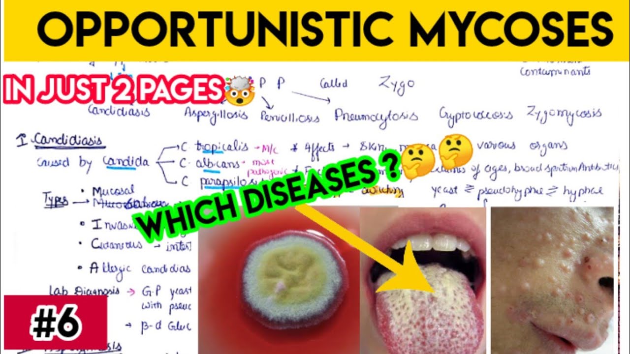
opportunistic mycoses - candidiasis aspergillosis zygomycosis in simplified way
Infecciones por hongos en la piel comunes, pero difíciles de detectar
5.0 / 5 (0 votes)